
- school Campus Bookshelves
- menu_book Bookshelves
- perm_media Learning Objects
- login Login
- how_to_reg Request Instructor Account
- hub Instructor Commons
- Download Page (PDF)
- Download Full Book (PDF)
- Periodic Table
- Physics Constants
- Scientific Calculator
- Reference & Cite
- Tools expand_more
- Readability
selected template will load here
This action is not available.

42.2: The Mechanism of Nerve Impulse Transmission
- Last updated
- Save as PDF
- Page ID 74335

Skills to Develop
- Describe the basis of the resting membrane potential
- Explain the stages of an action potential and how action potentials are propagated
- Explain the similarities and differences between chemical and electrical synapses
- Describe long-term potentiation and long-term depression
All functions performed by the nervous system—from a simple motor reflex to more advanced functions like making a memory or a decision—require neurons to communicate with one another. While humans use words and body language to communicate, neurons use electrical and chemical signals. Just like a person in a committee, one neuron usually receives and synthesizes messages from multiple other neurons before “making the decision” to send the message on to other neurons.
Nerve Impulse Transmission within a Neuron
For the nervous system to function, neurons must be able to send and receive signals. These signals are possible because each neuron has a charged cellular membrane (a voltage difference between the inside and the outside), and the charge of this membrane can change in response to neurotransmitter molecules released from other neurons and environmental stimuli. To understand how neurons communicate, one must first understand the basis of the baseline or ‘resting’ membrane charge.
Neuronal Charged Membranes
The lipid bilayer membrane that surrounds a neuron is impermeable to charged molecules or ions. To enter or exit the neuron, ions must pass through special proteins called ion channels that span the membrane. Ion channels have different configurations: open, closed, and inactive, as illustrated in Figure \(\PageIndex{1}\). Some ion channels need to be activated in order to open and allow ions to pass into or out of the cell. These ion channels are sensitive to the environment and can change their shape accordingly. Ion channels that change their structure in response to voltage changes are called voltage-gated ion channels. Voltage-gated ion channels regulate the relative concentrations of different ions inside and outside the cell. The difference in total charge between the inside and outside of the cell is called the membrane potential .

Link to Learning
This video discusses the basis of the resting membrane potential.
Resting Membrane Potential
A neuron at rest is negatively charged: the inside of a cell is approximately 70 millivolts more negative than the outside (−70 mV, note that this number varies by neuron type and by species). This voltage is called the resting membrane potential; it is caused by differences in the concentrations of ions inside and outside the cell. If the membrane were equally permeable to all ions, each type of ion would flow across the membrane and the system would reach equilibrium. Because ions cannot simply cross the membrane at will, there are different concentrations of several ions inside and outside the cell, as shown in the table below. The difference in the number of positively charged potassium ions (K + ) inside and outside the cell dominates the resting membrane potential (Figure \(\PageIndex{2}\)). When the membrane is at rest, K + ions accumulate inside the cell due to a net movement with the concentration gradient. The negative resting membrane potential is created and maintained by increasing the concentration of cations outside the cell (in the extracellular fluid) relative to inside the cell (in the cytoplasm). The negative charge within the cell is created by the cell membrane being more permeable to potassium ion movement than sodium ion movement. In neurons, potassium ions are maintained at high concentrations within the cell while sodium ions are maintained at high concentrations outside of the cell. The cell possesses potassium and sodium leakage channels that allow the two cations to diffuse down their concentration gradient. However, the neurons have far more potassium leakage channels than sodium leakage channels. Therefore, potassium diffuses out of the cell at a much faster rate than sodium leaks in. Because more cations are leaving the cell than are entering, this causes the interior of the cell to be negatively charged relative to the outside of the cell. The actions of the sodium potassium pump help to maintain the resting potential, once established. Recall that sodium potassium pumps brings two K + ions into the cell while removing three Na + ions per ATP consumed. As more cations are expelled from the cell than taken in, the inside of the cell remains negatively charged relative to the extracellular fluid. It should be noted that calcium ions (Cl – ) tend to accumulate outside of the cell because they are repelled by negatively-charged proteins within the cytoplasm.

Action Potential
A neuron can receive input from other neurons and, if this input is strong enough, send the signal to downstream neurons. Transmission of a signal between neurons is generally carried by a chemical called a neurotransmitter. Transmission of a signal within a neuron (from dendrite to axon terminal) is carried by a brief reversal of the resting membrane potential called an action potential . When neurotransmitter molecules bind to receptors located on a neuron’s dendrites, ion channels open. At excitatory synapses, this opening allows positive ions to enter the neuron and results in depolarization of the membrane—a decrease in the difference in voltage between the inside and outside of the neuron. A stimulus from a sensory cell or another neuron depolarizes the target neuron to its threshold potential (-55 mV). Na + channels in the axon hillock open, allowing positive ions to enter the cell (Figure \(\PageIndex{3}\) and Figure \(\PageIndex{4}\)). Once the sodium channels open, the neuron completely depolarizes to a membrane potential of about +40 mV. Action potentials are considered an "all-or nothing" event, in that, once the threshold potential is reached, the neuron always completely depolarizes. Once depolarization is complete, the cell must now "reset" its membrane voltage back to the resting potential. To accomplish this, the Na + channels close and cannot be opened. This begins the neuron's refractory period , in which it cannot produce another action potential because its sodium channels will not open. At the same time, voltage-gated K + channels open, allowing K + to leave the cell. As K + ions leave the cell, the membrane potential once again becomes negative. The diffusion of K + out of the cell actually hyperpolarizes the cell, in that the membrane potential becomes more negative than the cell's normal resting potential. At this point, the sodium channels will return to their resting state, meaning they are ready to open again if the membrane potential again exceeds the threshold potential. Eventually the extra K + ions diffuse out of the cell through the potassium leakage channels, bringing the cell from its hyperpolarized state, back to its resting membrane potential.
Art Connection

Potassium channel blockers, such as amiodarone and procainamide, which are used to treat abnormal electrical activity in the heart, called cardiac dysrhythmia, impede the movement of K + through voltage-gated K + channels. Which part of the action potential would you expect potassium channels to affect?

This video presents an overview of action potential.
Myelin and the Propagation of the Action Potential
For an action potential to communicate information to another neuron, it must travel along the axon and reach the axon terminals where it can initiate neurotransmitter release. The speed of conduction of an action potential along an axon is influenced by both the diameter of the axon and the axon’s resistance to current leak. Myelin acts as an insulator that prevents current from leaving the axon; this increases the speed of action potential conduction. In demyelinating diseases like multiple sclerosis, action potential conduction slows because current leaks from previously insulated axon areas. The nodes of Ranvier, illustrated in Figure \(\PageIndex{5}\) are gaps in the myelin sheath along the axon. These unmyelinated spaces are about one micrometer long and contain voltage gated Na + and K + channels. Flow of ions through these channels, particularly the Na + channels, regenerates the action potential over and over again along the axon. This ‘jumping’ of the action potential from one node to the next is called saltatory conduction . If nodes of Ranvier were not present along an axon, the action potential would propagate very slowly since Na + and K + channels would have to continuously regenerate action potentials at every point along the axon instead of at specific points. Nodes of Ranvier also save energy for the neuron since the channels only need to be present at the nodes and not along the entire axon.

Synaptic Transmission
The synapse or “gap” is the place where information is transmitted from one neuron to another. Synapses usually form between axon terminals and dendritic spines, but this is not universally true. There are also axon-to-axon, dendrite-to-dendrite, and axon-to-cell body synapses. The neuron transmitting the signal is called the presynaptic neuron, and the neuron receiving the signal is called the postsynaptic neuron. Note that these designations are relative to a particular synapse—most neurons are both presynaptic and postsynaptic. There are two types of synapses: chemical and electrical.
Chemical Synapse
When an action potential reaches the axon terminal it depolarizes the membrane and opens voltage-gated Na + channels. Na + ions enter the cell, further depolarizing the presynaptic membrane. This depolarization causes voltage-gated Ca 2+ channels to open. Calcium ions entering the cell initiate a signaling cascade that causes small membrane-bound vesicles, called synaptic vesicles , containing neurotransmitter molecules to fuse with the presynaptic membrane. Synaptic vesicles are shown in Figure \(\PageIndex{6}\), which is an image from a scanning electron microscope.

Fusion of a vesicle with the presynaptic membrane causes neurotransmitter to be released into the synaptic cleft , the extracellular space between the presynaptic and postsynaptic membranes, as illustrated in Figure \(\PageIndex{7}\). The neurotransmitter diffuses across the synaptic cleft and binds to receptor proteins on the postsynaptic membrane.

The binding of a specific neurotransmitter causes particular ion channels, in this case ligand-gated channels, on the postsynaptic membrane to open. Neurotransmitters can either have excitatory or inhibitory effects on the postsynaptic membrane, as detailed in the table below. For example, when acetylcholine is released at the synapse between a nerve and muscle (called the neuromuscular junction) by a presynaptic neuron, it causes postsynaptic Na + channels to open. Na + enters the postsynaptic cell and causes the postsynaptic membrane to depolarize. This depolarization is called an excitatory postsynaptic potential (EPSP) and makes the postsynaptic neuron more likely to fire an action potential. Release of neurotransmitter at inhibitory synapses causes inhibitory postsynaptic potentials (IPSPs) , a hyperpolarization of the presynaptic membrane. For example, when the neurotransmitter GABA (gamma-aminobutyric acid) is released from a presynaptic neuron, it binds to and opens Cl - channels. Cl - ions enter the cell and hyperpolarizes the membrane, making the neuron less likely to fire an action potential.
Once neurotransmission has occurred, the neurotransmitter must be removed from the synaptic cleft so the postsynaptic membrane can “reset” and be ready to receive another signal. This can be accomplished in three ways: the neurotransmitter can diffuse away from the synaptic cleft, it can be degraded by enzymes in the synaptic cleft, or it can be recycled (sometimes called reuptake) by the presynaptic neuron. Several drugs act at this step of neurotransmission. For example, some drugs that are given to Alzheimer’s patients work by inhibiting acetylcholinesterase, the enzyme that degrades acetylcholine. This inhibition of the enzyme essentially increases neurotransmission at synapses that release acetylcholine. Once released, the acetylcholine stays in the cleft and can continually bind and unbind to postsynaptic receptors.
Electrical Synapse
While electrical synapses are fewer in number than chemical synapses, they are found in all nervous systems and play important and unique roles. The mode of neurotransmission in electrical synapses is quite different from that in chemical synapses. In an electrical synapse, the presynaptic and postsynaptic membranes are very close together and are actually physically connected by channel proteins forming gap junctions. Gap junctions allow current to pass directly from one cell to the next. In addition to the ions that carry this current, other molecules, such as ATP, can diffuse through the large gap junction pores.
There are key differences between chemical and electrical synapses. Because chemical synapses depend on the release of neurotransmitter molecules from synaptic vesicles to pass on their signal, there is an approximately one millisecond delay between when the axon potential reaches the presynaptic terminal and when the neurotransmitter leads to opening of postsynaptic ion channels. Additionally, this signaling is unidirectional. Signaling in electrical synapses, in contrast, is virtually instantaneous (which is important for synapses involved in key reflexes), and some electrical synapses are bidirectional. Electrical synapses are also more reliable as they are less likely to be blocked, and they are important for synchronizing the electrical activity of a group of neurons. For example, electrical synapses in the thalamus are thought to regulate slow-wave sleep, and disruption of these synapses can cause seizures.
Signal Summation
Sometimes a single EPSP is strong enough to induce an action potential in the postsynaptic neuron, but often multiple presynaptic inputs must create EPSPs around the same time for the postsynaptic neuron to be sufficiently depolarized to fire an action potential. This process is called summation and occurs at the axon hillock, as illustrated in Figure \(\PageIndex{8}\). Additionally, one neuron often has inputs from many presynaptic neurons—some excitatory and some inhibitory—so IPSPs can cancel out EPSPs and vice versa. It is the net change in postsynaptic membrane voltage that determines whether the postsynaptic cell has reached its threshold of excitation needed to fire an action potential. Together, synaptic summation and the threshold for excitation act as a filter so that random “noise” in the system is not transmitted as important information.

Everyday Connection: Brain-computer interface
Amyotrophic lateral sclerosis (ALS, also called Lou Gehrig’s Disease) is a neurological disease characterized by the degeneration of the motor neurons that control voluntary movements. The disease begins with muscle weakening and lack of coordination and eventually destroys the neurons that control speech, breathing, and swallowing; in the end, the disease can lead to paralysis. At that point, patients require assistance from machines to be able to breathe and to communicate. Several special technologies have been developed to allow “locked-in” patients to communicate with the rest of the world. One technology, for example, allows patients to type out sentences by twitching their cheek. These sentences can then be read aloud by a computer.
A relatively new line of research for helping paralyzed patients, including those with ALS, to communicate and retain a degree of self-sufficiency is called brain-computer interface (BCI) technology and is illustrated in Figure \(\PageIndex{9}\). This technology sounds like something out of science fiction: it allows paralyzed patients to control a computer using only their thoughts. There are several forms of BCI. Some forms use EEG recordings from electrodes taped onto the skull. These recordings contain information from large populations of neurons that can be decoded by a computer. Other forms of BCI require the implantation of an array of electrodes smaller than a postage stamp in the arm and hand area of the motor cortex. This form of BCI, while more invasive, is very powerful as each electrode can record actual action potentials from one or more neurons. These signals are then sent to a computer, which has been trained to decode the signal and feed it to a tool—such as a cursor on a computer screen. This means that a patient with ALS can use e-mail, read the Internet, and communicate with others by thinking of moving his or her hand or arm (even though the paralyzed patient cannot make that bodily movement). Recent advances have allowed a paralyzed locked-in patient who suffered a stroke 15 years ago to control a robotic arm and even to feed herself coffee using BCI technology.
Despite the amazing advancements in BCI technology, it also has limitations. The technology can require many hours of training and long periods of intense concentration for the patient; it can also require brain surgery to implant the devices.

Watch this video in which a paralyzed woman use a brain-controlled robotic arm to bring a drink to her mouth, among other images of brain-computer interface technology in action.
Synaptic Plasticity
Synapses are not static structures. They can be weakened or strengthened. They can be broken, and new synapses can be made. Synaptic plasticity allows for these changes, which are all needed for a functioning nervous system. In fact, synaptic plasticity is the basis of learning and memory. Two processes in particular, long-term potentiation (LTP) and long-term depression (LTD) are important forms of synaptic plasticity that occur in synapses in the hippocampus, a brain region that is involved in storing memories.
Long-term Potentiation (LTP)
Long-term potentiation (LTP) is a persistent strengthening of a synaptic connection. LTP is based on the Hebbian principle: cells that fire together wire together. There are various mechanisms, none fully understood, behind the synaptic strengthening seen with LTP. One known mechanism involves a type of postsynaptic glutamate receptor, called NMDA (N-Methyl-D-aspartate) receptors, shown in Figure \(\PageIndex{10}\). These receptors are normally blocked by magnesium ions; however, when the postsynaptic neuron is depolarized by multiple presynaptic inputs in quick succession (either from one neuron or multiple neurons), the magnesium ions are forced out allowing Ca ions to pass into the postsynaptic cell. Next, Ca 2+ ions entering the cell initiate a signaling cascade that causes a different type of glutamate receptor, called AMPA (α-amino-3-hydroxy-5-methyl-4-isoxazolepropionic acid) receptors, to be inserted into the postsynaptic membrane, since activated AMPA receptors allow positive ions to enter the cell. So, the next time glutamate is released from the presynaptic membrane, it will have a larger excitatory effect (EPSP) on the postsynaptic cell because the binding of glutamate to these AMPA receptors will allow more positive ions into the cell. The insertion of additional AMPA receptors strengthens the synapse and means that the postsynaptic neuron is more likely to fire in response to presynaptic neurotransmitter release. Some drugs of abuse co-opt the LTP pathway, and this synaptic strengthening can lead to addiction.
Long-term Depression (LTD)
Long-term depression (LTD) is essentially the reverse of LTP: it is a long-term weakening of a synaptic connection. One mechanism known to cause LTD also involves AMPA receptors. In this situation, calcium that enters through NMDA receptors initiates a different signaling cascade, which results in the removal of AMPA receptors from the postsynaptic membrane, as illustrated in Figure \(\PageIndex{10}\). The decrease in AMPA receptors in the membrane makes the postsynaptic neuron less responsive to glutamate released from the presynaptic neuron. While it may seem counterintuitive, LTD may be just as important for learning and memory as LTP. The weakening and pruning of unused synapses allows for unimportant connections to be lost and makes the synapses that have undergone LTP that much stronger by comparison.

Neurons have charged membranes because there are different concentrations of ions inside and outside of the cell. Voltage-gated ion channels control the movement of ions into and out of a neuron. When a neuronal membrane is depolarized to at least the threshold of excitation, an action potential is fired. The action potential is then propagated along a myelinated axon to the axon terminals. In a chemical synapse, the action potential causes release of neurotransmitter molecules into the synaptic cleft. Through binding to postsynaptic receptors, the neurotransmitter can cause excitatory or inhibitory postsynaptic potentials by depolarizing or hyperpolarizing, respectively, the postsynaptic membrane. In electrical synapses, the action potential is directly communicated to the postsynaptic cell through gap junctions—large channel proteins that connect the pre-and postsynaptic membranes. Synapses are not static structures and can be strengthened and weakened. Two mechanisms of synaptic plasticity are long-term potentiation and long-term depression.
Art Connections
Figure \(\PageIndex{3}\): Potassium channel blockers, such as amiodarone and procainamide, which are used to treat abnormal electrical activity in the heart, called cardiac dysrhythmia, impede the movement of K + through voltage-gated K + channels. Which part of the action potential would you expect potassium channels to affect?
Potassium channel blockers slow the repolarization phase, but have no effect on depolarization.
- Anatomy and Physiology
- Study Guides
- Transmission of Nerve Impulses
- Quiz: What is Anatomy and Physiology?
- Atoms, Molecules, Ions, and Bonds
- Quiz: Atoms, Molecules, Ions, and Bonds
- Inorganic Compounds
- Quiz: Inorganic Compounds
- Organic Molecules
- What Is Anatomy and Physiology?
- Quiz: Organic Molecules
- Chemical Reactions in Metabolic Processes
- Quiz: Chemical Reactions in Metabolic Processes
- Quiz: The Cell and Its Membrane
- Cell Junctions
- Quiz: Cell Junctions
- Movement of Substances
- Quiz: Movement of Substances
- Cell Division
- The Cell and Its Membrane
- Quiz: Cell Division
- Epithelial Tissue
- Quiz: Epithelial Tissue
- Connective Tissue
- Quiz: Connective Tissue
- Nervous Tissue
- Introduction to Tissues
- Quiz: Nervous Tissue
- Muscle Tissue
- Quiz: Muscle Tissue
- Quiz: The Skin and Its Functions
- The Epidermis
- Quiz: The Epidermis
- Quiz: The Dermis
- The Hypodermis
- The Skin and Its Functions
- Quiz: The Hypodermis
- Accessory Organs of the Skin
- Quiz: Accessory Organs of the Skin
- Quiz: Types of Bones
- Bone Structure
- Quiz: Bone Structure
- Bone Development
- Quiz: Bone Development
- Bone Growth
- Functions of Bones
- Quiz: Functions of Bones
- Types of Bones
- Quiz: Bone Growth
- Bone Homeostasis
- Quiz: Bone Homeostasis
- Surface Features of Bones
- Quiz: Surface Features of Bones
- Quiz: Skull: Cranium and Facial Bones
- Quiz: Hyoid Bone
- Vertebral Column
- Quiz: Vertebral Column
- Organization of the Skeleton
- Quiz: Organization of the Skeleton
- Skull: Cranium and Facial Bones
- Quiz: Thorax
- Pectoral Girdle
- Quiz: Pectoral Girdle
- Quiz: Upper Limb
- Pelvic Girdle
- Quiz: Pelvic Girdle
- Quiz: Lower Limb
- Classifying Joints
- Quiz: Classifying Joints
- Quiz: Types of Muscles
- Connective Tissue Associated with Muscle Tissue
- Quiz: Connective Tissue Associated with Muscle Tissue
- Structure of Skeletal Muscle
- Quiz: Structure of Skeletal Muscle
- Muscle Contraction
- Types of Muscles
- Quiz: Muscle Contraction
- Muscle Metabolism
- Structure of Cardiac and Smooth Muscle
- Quiz: Structure of Cardiac and Smooth Muscle
- Quiz: Skeletal Muscle Actions
- Names of Skeletal Muscles
- Quiz: Names of Skeletal Muscles
- Muscle Size and Arrangement of Muscle Fascicles
- Quiz: Muscle Size and Arrangement of Muscle Fascicles
- Major Skeletal Muscles
- Skeletal Muscle Actions
- Quiz: Major Skeletal Muscles
- Quiz: Neuroglia
- Myelination
- Quiz: Myelination
- Quiz: Neurons
- Quiz: Transmission of Nerve Impulses
- The Synapse
- Quiz: The Synapse
- Nervous System Terminology
- Quiz: Nervous System Terminology
- Quiz: The Brain
- The Ventricles and Cerebrospinal Fluid
- Nervous System Organization
- Quiz: Nervous System Organization
- Quiz: The Ventricles and Cerebrospinal Fluid
- The Meninges
- Quiz: The Meninges
- The Blood-Brain Barrier
- Quiz: The Blood-Brain Barrier
- Cranial Nerves
- Quiz: Cranial Nerves
- The Spinal Cord
- Quiz: The Spinal Cord
- Spinal Nerves
- Quiz: Spinal Nerves
- Quiz: Reflexes
- The Autonomic Nervous System
- Quiz: The Autonomic Nervous System
- Quiz: Sensory Receptors
- The Somatic Senses
- Quiz: The Somatic Senses
- Quiz: Vision
- Sensory Receptors
- Quiz: Hearing
- Equilibrium
- Quiz: Equilibrium
- Quiz: Smell
- Quiz: Taste
- Quiz: The Hypothalamus and Pituitary Glands
- Endocrine Organs and Tissues
- Quiz: Endocrine Organs and Tissues
- Antagonistic Hormones
- Quiz: Antagonistic Hormones
- Quiz: Hormones
- The Hypothalamus and Pituitary Glands
- Quiz: The Blood
- Blood Formation
- Quiz: Blood Formation
- Quiz: Hemostasis
- Blood Groups
- Functions of the Cardiovascular System
- Quiz: Functions of the Cardiovascular System
- Quiz: Blood Groups
- Circulatory Pathways
- Quiz: Circulatory Pathways
- Quiz: The Heart
- Cardiac Conduction
- Cardiac Muscle Contraction
- Electrocardiogram
- The Cardiac Cycle
- Cardiac Output
- Blood Vessels
- Blood Pressure
- Control of Blood Pressure
- Blood Vessels of the Body
- Lymphatic Vessels
- Quiz: Lymphatic Vessels
- Lymphoid Cells
- Quiz: Lymphoid Cells
- Lymphatic Tissues and Organs
- Lymphatic System Components
- Quiz: Lymphatic System Components
- Quiz: Lymphatic Tissues and Organs
- Nonspecific Barriers
- Quiz: Nonspecific Barriers
- Nonspecific Defenses
- Quiz: Nonspecific Defenses
- Specific Defense (The Immune System)
- Protecting Your Body
- Quiz: Specific Defense (The Immune System)
- Major Histocompatibility Complex
- Quiz: Major Histocompatibility Complex
- Lymphocytes
- Quiz: Lymphocytes
- Quiz: Antibodies
- Costimulation
- Quiz: Costimulation
- Humoral and Cell-Mediated Immune Responses
- Quiz: Humoral and Cell-Mediated Immune Responses
- Supplements to the Immune Response
- Quiz: Supplements to the Immune Response
- Quiz: Structure of the Respiratory System
- Quiz: Lungs
- Mechanics of Breathing
- Quiz: Mechanics of Breathing
- Function of the Respiratory System
- Lung Volumes and Capacities
- Quiz: Function of the Respiratory System
- Structure of the Respiratory System
- Quiz: Lung Volumes and Capacities
- Gas Exchange
- Quiz: Gas Exchange
- Gas Transport
- Quiz: Gas Transport
- Control of Respiration
- Quiz: Control of Respiration
- Quiz: Structure of the Digestive Tract Wall
- Digestive Enzymes
- Quiz: Digestive Enzymes
- Quiz: The Mouth
- Function of the Digestive System
- Quiz: Function of the Digestive System
- Structure of the Digestive Tract Wall
- The Pharynx
- The Esophagus
- Quiz: The Esophagus
- Deglutition (Swallowing)
- Quiz: Deglutition (Swallowing)
- The Stomach
- Quiz: The Stomach
- The Small Intestine
- Quiz: The Small Intestine
- Large Intestine
- Quiz: Large Intestine
- The Pancreas
- Quiz: The Pancreas
- The Liver and Gallbladder
- Quiz: The Liver and Gallbladder
- Regulation of Digestion
- Quiz: Regulation of Digestion
- Quiz: Regulation of Urine Concentration
- Quiz: Ureters
- Urinary Bladder
- Quiz: Urinary Bladder
- Anatomy of the Kidneys
- Quiz: Anatomy of the Kidneys
- Regulation of Urine Concentration
- Quiz: Urethra
- Quiz: What Is Reproduction?
- The Male Reproductive System
- Quiz: The Male Reproductive System
- The Female Reproduction System
- Quiz: The Female Reproduction System
- What Is Reproduction?
- Anatomy and Physiology Quizzes
Polarization is established by maintaining an excess of sodium ions (Na + ) on the outside and an excess of potassium ions (K + ) on the inside. A certain amount of Na + and K + is always leaking across the membrane through leakage channels, but Na + /K + pumps in the membrane actively restore the ions to the appropriate side.
The main contribution to the resting membrane potential (a polarized nerve) is the difference in permeability of the resting membrane to potassium ions versus sodium ions. The resting membrane is much more permeable to potassium ions than to sodium ions resulting in slightly more net potassium ion diffusion (from the inside of the neuron to the outside) than sodium ion diffusion (from the outside of the neuron to the inside) causing the slight difference in polarity right along the membrane of the axon.
Other ions, such as large, negatively charged proteins and nucleic acids, reside within the cell. It is these large, negatively charged ions that contribute to the overall negative charge on the inside of the cell membrane as compared to the outside.
In addition to crossing the membrane through leakage channels, ions may cross through gated channels. Gated channels open in response to neurotransmitters, changes in membrane potential, or other stimuli.
The following events characterize the transmission of a nerve impulse (see Figure 1):
- Resting potential. The resting potential describes the unstimulated, polarized state of a neuron (at about –70 millivolts).
- Graded potential. A graded potential is a change in the resting potential of the plasma membrane in the response to a stimulus. A graded potential occurs when the stimulus causes Na + or K + gated channels to open. If Na + channels open, positive sodium ions enter, and the membrane depolarizes (becomes more positive). If the stimulus opens K + channels, then positive potassium ions exit across the membrane and the membrane hyperpolarizes (becomes more negative). A graded potential is a local event that does not travel far from its origin. Graded potentials occur in cell bodies and dendrites. Light, heat, mechanical pressure, and chemicals, such as neurotransmitters, are examples of stimuli that may generate a graded potential (depending upon the neuron).
Figure 1.Events that characterize the transmission of a nerve impulse.

The following four steps describe the initiation of an impulse to the “resetting” of a neuron to prepare for a second stimulation:
- Action potential. Unlike a graded potential, an action potential is capable of traveling long distances. If a depolarizing graded potential is sufficiently large, Na + channels in the trigger zone open. In response, Na + on the outside of the membrane becomes depolarized (as in a graded potential). If the stimulus is strong enough—that is, if it is above a certain threshold level—additional Na + gates open, increasing the flow of Na + even more, causing an action potential, or complete depolarization (from –70 to about +30 millivolts). This in turn stimulates neighboring Na + gates, farther down the axon, to open. In this manner, the action potential travels down the length of the axon as opened Na + gates stimulate neighboring Na + gates to open. The action potential is an all‐or‐nothing event: When the stimulus fails to produce depolarization that exceeds the threshold value, no action potential results, but when threshold potential is exceeded, complete depolarization occurs.
- Repolarization. In response to the inflow of Na + , K + channels open, this time allowing K + on the inside to rush out of the cell. The movement of K + out of the cell causes repolarization by restoring the original membrane polarization. Unlike the resting potential, however, in repolarization the K + are on the outside and the Na + are on the inside. Soon after the K + gates open, the Na + gates close.
- Hyperpolarization. By the time the K + channels close, more K + have moved out of the cell than is actually necessary to establish the original polarized potential. Thus, the membrane becomes hyperpolarized (about –80 millivolts).
- Refractory period. With the passage of the action potential, the cell membrane is in an unusual state of affairs. The membrane is polarized, but the Na + and K + are on the wrong sides of the membrane. During this refractory period, the axon will not respond to a new stimulus. To reestablish the original distribution of these ions, the Na + and K + are returned to their resting potential location by Na + /K + pumps in the cell membrane. Once these ions are completely returned to their resting potential location, the neuron is ready for another stimulus.
Previous Quiz: Myelination
Next Neurons
- Online Quizzes for CliffsNotes Anatomy and Physiology QuickReview, 2nd Edition
Want to create or adapt books like this? Learn more about how Pressbooks supports open publishing practices.
8.4 Nerve Impulses
Created by CK-12 Foundation/Adapted by Christine Miller
When Lightning Strikes
This amazing cloud-to-surface lightning occurred when a difference in electrical charge built up in a cloud relative to the ground. When the buildup of charge was great enough, a sudden discharge of electricity occurred. A nerve impulse is similar to a lightning strike. Both a nerve impulse and a lightning strike occur because of differences in electrical charge, and both result in an electric current.
Generating Nerve Impulses
A nerve impulse , like a lightning strike, is an electrical phenomenon. A nerve impulse occurs because of a difference in electrical charge across the plasma membrane of a neuron. How does this difference in electrical charge come about? The answer involves ions , which are electrically-charged atoms or molecules .
Resting Potential
When a neuron is not actively transmitting a nerve impulse, it is in a resting state, ready to transmit a nerve impulse. During the resting state, the sodium-potassium pump maintains a difference in charge across the cell membrane of the neuron. The sodium-potassium pump is a mechanism of active transport that moves sodium ions (Na+) out of cells and potassium ions (K+) into cells. The sodium-potassium pump moves both ions from areas of lower to higher concentration, using energy in ATP and carrier proteins in the cell membrane. The video below, “Sodium Potassium Pump” by Amoeba Sisters, describes in greater detail how the sodium-potassium pump works. Sodium is the principal ion in the fluid outside of cells, and potassium is the principal ion in the fluid inside of cells. These differences in concentration create an electrical gradient across the cell membrane, called resting potential . Tightly controlling membrane resting potential is critical for the transmission of nerve impulses.
Sodium Potassium Pump, Amoeba Sisters, 2020.
Action Potential
A nerve impulse is a sudden reversal of the electrical gradient across the plasma membrane of a resting neuron. The reversal of charge is called an action potential . It begins when the neuron receives a chemical signal from another cell or some other type of stimulus . If the stimulus is strong enough to reach threshold , an action potential will take place is a cascade along the axon.
This reversal of charges ripples down the axon of the neuron very rapidly as an electric current, which is illustrated in the diagram below (Figure 8.4.2). A nerve impulse is an all-or-nothing response depending on if the stimulus input was strong enough to reach threshold. If a neuron responds at all, it responds completely. A greater stimulation does not produce a stronger impulse.
In neurons with a myelin sheath on their axon, ions flow across the membrane only at the nodes between sections of myelin. As a result, the action potential appears to jump along the axon membrane from node to node, rather than spreading smoothly along the entire membrane. This increases the speed at which the action potential travels.
Transmitting Nerve Impulses
The place where an axon terminal meets another cell is called a synapse . This is where the transmission of a nerve impulse to another cell occurs. The cell that sends the nerve impulse is called the presynaptic cell , and the cell that receives the nerve impulse is called the postsynaptic cell .
Some synapses are purely electrical and make direct electrical connections between neurons. Most synapses, however, are chemical synapses. Transmission of nerve impulses across chemical synapses is more complex.
Chemical Synapses
At a chemical synapse, both the presynaptic and postsynaptic areas of the cells are full of molecular machinery that is involved in the transmission of nerve impulses. As shown in Figure 8.4.3, the presynaptic area contains many tiny spherical vessels called synaptic vesicles that are packed with chemicals called neurotransmitters . When an action potential reaches the axon terminal of the presynaptic cell, it opens channels that allow calcium to enter the terminal. Calcium causes synaptic vesicles to fuse with the membrane, releasing their contents into the narrow space between the presynaptic and postsynaptic membranes. This area is called the synaptic cleft . The neurotransmitter molecules travel across the synaptic cleft and bind to receptors , which are proteins embedded in the membrane of the postsynaptic cell.
Neurotransmitters and Receptors
There are more than a hundred known neurotransmitters, and more than one type of neurotransmitter may be released at a given synapse by a presynaptic cell. For example, it is common for a faster-acting neurotransmitter to be released, along with a slower-acting neurotransmitter. Many neurotransmitters also have multiple types of receptors to which they can bind. Receptors, in turn, can be divided into two general groups: chemically gated ion channels and second messenger systems.
- When a chemically gated ion channel is activated, it forms a passage that allows specific types of ions to flow across the cell membrane. Depending on the type of ion, the effect on the target cell may be excitatory or inhibitory .
- When a second messenger system is activated, it starts a cascade of molecular interactions inside the target cell. This may ultimately produce a wide variety of complex effects, such as increasing or decreasing the sensitivity of the cell to stimuli, or even altering gene transcription.
The effect of a neurotransmitter on a postsynaptic cell depends mainly on the type of receptors that it activates, making it possible for a particular neurotransmitter to have different effects on various target cells. A neurotransmitter might excite one set of target cells, inhibit others, and have complex modulatory effects on still others, depending on the type of receptors. However, some neurotransmitters have relatively consistent effects on other cells. Consider the two most widely used neurotransmitters, glutamate and GABA (gamma-aminobutyric acid). Glutamate receptors are either excitatory or modulatory in their effects, whereas GABA receptors are all inhibitory in their effects in adults.
Problems with neurotransmitters or their receptors can cause neurological disorders. The disease myasthenia gravis , for example, is caused by antibodies from the immune system blocking receptors for the neurotransmitter acetylcholine in postsynaptic muscle cells. This inhibits the effects of acetylcholine on muscle contractions, producing symptoms, such as muscle weakness and excessive fatigue during simple activities. Some mental illnesses (including depression ) are caused, at least in part, by imbalances of certain neurotransmitters in the brain. One of the neurotransmitters involved in depression is thought to be serotonin , which normally helps regulate mood, among many other functions. Some antidepressant drugs are thought to help alleviate depression in many patients by normalizing the activity of serotonin in the brain.
8.4 Summary
- A nerve impulse is an electrical phenomenon that occurs because of a difference in electrical charge across the plasma membrane of a neuron.
- The sodium-potassium pump maintains an electrical gradient across the plasma membrane of a neuron when it is not actively transmitting a nerve impulse. This gradient is called the resting potential of the neuron.
- An action potential is a sudden reversal of the electrical gradient across the plasma membrane of a resting neuron. It begins when the neuron receives a chemical signal from another cell or some other type of stimulus. The action potential travels rapidly down the neuron’s axon as an electric current and occurs in three stages: Depolarization, Repolarization and Recovery.
- A nerve impulse is transmitted to another cell at either an electrical or a chemical synapse . At a chemical synapse, neurotransmitter chemicals are released from the presynaptic cell into the synaptic cleft between cells. The chemicals travel across the cleft to the postsynaptic cell and bind to receptors embedded in its membrane.
- There are many different types of neurotransmitters. Their effects on the postsynaptic cell generally depend on the type of receptor they bind to. The effects may be excitatory, inhibitory, or modulatory in more complex ways. Both physical and mental disorders may occur if there are problems with neurotransmitters or their receptors.
8.4 Review Questions
- Define nerve impulse.
- What is the resting potential of a neuron, and how is it maintained?
- Explain how and why an action potential occurs.
- Outline how a signal is transmitted from a presynaptic cell to a postsynaptic cell at a chemical synapse.
- What generally determines the effects of a neurotransmitter on a postsynaptic cell?
- Identify three general types of effects that neurotransmitters may have on postsynaptic cells.
- Explain how an electrical signal in a presynaptic neuron causes the transmission of a chemical signal at the synapse.
- The flow of which type of ion into a neuron results in an action potential? How do these ions get into the cell? What does this flow of ions do to the relative charge inside the neuron compared to the outside?
- Name three neurotransmitters.
8.4 Explore More
Action Potentials, Teacher’s Pet, 2018.
TED Ed| What is depression? – Helen M. Farrell, Parta Learning, 2017.
5 Weird Involuntary Behaviors Explained!, It’s Okay To Be Smart, 2015.
Attributions
Figure 8.4.1
Lightening/ Purple Lightning, Dee Why by Jeremy Bishop on Unsplash is used under the Unsplash License (https://unsplash.com/license).
Figure 8.4.2
Action Potential by CNX OpenStax, Biology on Wikimedia Commons is used under a CC BY 4.0 (https://creativecommons.org/licenses/by/4.0/deed.en) license.
Figure 8.4.3
Chemical_synapse_schema_cropped by Looie496 created file (adapted from original from US National Institutes of Health, National Institute on Aging) is in the public domain (https://en.wikipedia.org/wiki/Public_domain).
Amoeba Sisters. (2020, January 29). Sodium potassium pump. YouTube. https://www.youtube.com/watch?v=7NY6XdPBhxo&feature=youtu.be
CNX OpenStax. (2016, May 27) Figure 4 The action potential is conducted down the axon as the axon membrane depolarizes, then repolarizes [digital image]. In Open Stax, Biology (Section 35.2). OpenStax CNX. https://cnx.org/contents/[email protected]:cs_Pb-GW@6/How-Neurons-Communicate
It’s Okay To Be Smart. (2015, January 26). 5 Weird involuntary behaviors explained! YouTube. https://www.youtube.com/watch?v=ZE8sRMZ5BCA&feature=youtu.be
Mayo Clinic Staff. (n.d.). Depression (major depressive disorder) [online article]. MayoClinic.org. https://www.mayoclinic.org/diseases-conditions/depression/symptoms-causes/syc-20356007
Mayo Clinic Staff. (n.d.). Myasthenia gravis [online article]. MayoClinic.org. https://www.mayoclinic.org/diseases-conditions/myasthenia-gravis/symptoms-causes/syc-20352036
National Institute on Aging. (2006, April 8). Alzheimers disease: Unraveling the mystery. National Institutes of Health. https://www.nia.nih.gov/ ( archived version )
Parta Learning. (2017, December 8). TED Ed| What is depression? – Helen M. Farrell. YouTube. https://www.youtube.com/watch?v=rBcU_apy0h8&t=291s
Teacher’s Pet. (2018, August 26). Action potentials. YouTube. https://www.youtube.com/watch?v=FEHNIELPb0s&feature=youtu.be
A signal transmitted along a nerve fiber.
An atom or molecule with a net electric charge due to the loss or gain of one or more electrons.
The smallest particle of an element that still has the properties of that element.
A molecule is an electrically neutral group of two or more atoms held together by chemical bonds.
A functional unit of the nervous system that transmits nerve impulses; also called a nerve cell.
A solute pump that pumps potassium into cells while pumping sodium out of cells, both against their concentration gradients. This pumping is active and occurs at the ratio of 2 potassium for every 3 calcium.
The semipermeable membrane surrounding the cytoplasm of a cell.
The movement of ions or molecules across a cell membrane into a region of higher concentration, assisted by enzymes and requiring energy.
A complex organic chemical that provides energy to drive many processes in living cells, e.g. muscle contraction, nerve impulse propagation, and chemical synthesis. Found in all forms of life, ATP is often referred to as the "molecular unit of currency" of intracellular energy transfer.
The difference in electrical charge across the plasma membrane of a neuron that is not actively transmitting a nerve impulse.
Reversal of electrical charge across the membrane of a resting neuron that travels down the axon of the neuron as a nerve impulse.
Something that triggers a behavior or other response.
The critical level to which a membrane potential must be depolarized to initiate an action potential.
The place where the axon terminal of a neuron transmits a chemical or electrical signal to another cell.
The cell that sends the nerve impulse.
The cell that receives the nerve impulse.
These membrane-bound organelles store various neurotransmitters that are released at the synapse. The release is regulated by a voltage-dependent calcium channel. Vesicles are essential for propagating nerve impulses between neurons and are constantly recreated by the cell.
A type of chemical that transmits signals from the axon of a neuron to another cell across a synapse.
A space that separates two neurons. It forms a junction between two or more neurons and helps nerve impulse pass from one neuron to the other.
A protein on a cell membrane or inside of a cell that binds with a hormone, neurotransmitter, or other chemical signal to produce a response.
A neurotransmitter that will have excitatory effects on the neuron, meaning it will increase the likelihood that a neuron will fire an action potential.
A neurotransmitter that decreases the likelihood that a neuron will fire an action potential.
A chemical that nerve cells use to send signals to other cells. It is by a wide margin the most abundant excitatory neurotransmitter in the vertebrate nervous system.
A naturally occurring amino acid that works as a neurotransmitter in your brain. Neurotransmitters function as chemical messengers. GABA is considered an inhibitory neurotransmitter because it blocks, or inhibits, certain brain signals and decreases activity in your nervous system.
An antibody, also known as an immunoglobulin, is a large, Y-shaped protein produced mainly by plasma cells that is used by the immune system to neutralize pathogens such as pathogenic bacteria and viruses.
An organic chemical that functions in the brain and body of many types of animals (and humans) as a neurotransmitter—a chemical message released by nerve cells to send signals to other cells, such as neurons, muscle cells and gland cells.
A neurotransmitter. It has a popular image as a contributor to feelings of well-being and happiness, though its actual biological function is complex and multifaceted, modulating cognition, reward, learning, memory, and numerous physiological processes such as vomiting and vasoconstriction.
Human Biology Copyright © 2020 by Christine Miller is licensed under a Creative Commons Attribution-NonCommercial 4.0 International License , except where otherwise noted.
Share This Book

Want to create or adapt books like this? Learn more about how Pressbooks supports open publishing practices.
80 8.4 Nerve Impulses
Created by CK-12 Foundation/Adapted by Christine Miller

When Lightning Strikes
This amazing cloud-to-surface lightning occurred when a difference in electrical charge built up in a cloud relative to the ground. When the buildup of charge was great enough, a sudden discharge of electricity occurred. A nerve impulse is similar to a lightning strike. Both a nerve impulse and a lightning strike occur because of differences in electrical charge, and both result in an electric current.
Generating Nerve Impulses
A nerve impulse , like a lightning strike, is an electrical phenomenon. A nerve impulse occurs because of a difference in electrical charge across the plasma membrane of a neuron. How does this difference in electrical charge come about? The answer involves ions , which are electrically-charged atoms or molecules .
Resting Potential
When a neuron is not actively transmitting a nerve impulse, it is in a resting state, ready to transmit a nerve impulse. During the resting state, the sodium-potassium pump maintains a difference in charge across the cell membrane of the neuron. The sodium-potassium pump is a mechanism of active transport that moves sodium ions (Na+) out of cells and potassium ions (K+) into cells. The sodium-potassium pump moves both ions from areas of lower to higher concentration, using energy in ATP and carrier proteins in the cell membrane. The video below, “Sodium Potassium Pump” by Amoeba Sisters, describes in greater detail how the sodium-potassium pump works. Sodium is the principal ion in the fluid outside of cells, and potassium is the principal ion in the fluid inside of cells. These differences in concentration create an electrical gradient across the cell membrane, called resting potential . Tightly controlling membrane resting potential is critical for the transmission of nerve impulses.
Sodium Potassium Pump, Amoeba Sisters, 2020.
Action Potential
A nerve impulse is a sudden reversal of the electrical gradient across the plasma membrane of a resting neuron. The reversal of charge is called an action potential . It begins when the neuron receives a chemical signal from another cell or some other type of stimulus . If the stimulus is strong enough to reach threshold , an action potential will take place is a cascade along the axon.
This reversal of charges ripples down the axon of the neuron very rapidly as an electric current, which is illustrated in the diagram below (Figure 8.4.2). A nerve impulse is an all-or-nothing response depending on if the stimulus input was strong enough to reach threshold. If a neuron responds at all, it responds completely. A greater stimulation does not produce a stronger impulse.

In neurons with a myelin sheath on their axon, ions flow across the membrane only at the nodes between sections of myelin. As a result, the action potential appears to jump along the axon membrane from node to node, rather than spreading smoothly along the entire membrane. This increases the speed at which the action potential travels.
Transmitting Nerve Impulses
The place where an axon terminal meets another cell is called a synapse . This is where the transmission of a nerve impulse to another cell occurs. The cell that sends the nerve impulse is called the presynaptic cell , and the cell that receives the nerve impulse is called the postsynaptic cell .
Some synapses are purely electrical and make direct electrical connections between neurons. Most synapses, however, are chemical synapses. Transmission of nerve impulses across chemical synapses is more complex.
Chemical Synapses
At a chemical synapse, both the presynaptic and postsynaptic areas of the cells are full of molecular machinery that is involved in the transmission of nerve impulses. As shown in Figure 8.4.3, the presynaptic area contains many tiny spherical vessels called synaptic vesicles that are packed with chemicals called neurotransmitters . When an action potential reaches the axon terminal of the presynaptic cell, it opens channels that allow calcium to enter the terminal. Calcium causes synaptic vesicles to fuse with the membrane, releasing their contents into the narrow space between the presynaptic and postsynaptic membranes. This area is called the synaptic cleft . The neurotransmitter molecules travel across the synaptic cleft and bind to receptors , which are proteins embedded in the membrane of the postsynaptic cell.

Neurotransmitters and Receptors
There are more than a hundred known neurotransmitters, and more than one type of neurotransmitter may be released at a given synapse by a presynaptic cell. For example, it is common for a faster-acting neurotransmitter to be released, along with a slower-acting neurotransmitter. Many neurotransmitters also have multiple types of receptors to which they can bind. Receptors, in turn, can be divided into two general groups: chemically gated ion channels and second messenger systems.
- When a chemically gated ion channel is activated, it forms a passage that allows specific types of ions to flow across the cell membrane. Depending on the type of ion, the effect on the target cell may be excitatory or inhibitory .
- When a second messenger system is activated, it starts a cascade of molecular interactions inside the target cell. This may ultimately produce a wide variety of complex effects, such as increasing or decreasing the sensitivity of the cell to stimuli, or even altering gene transcription.
The effect of a neurotransmitter on a postsynaptic cell depends mainly on the type of receptors that it activates, making it possible for a particular neurotransmitter to have different effects on various target cells. A neurotransmitter might excite one set of target cells, inhibit others, and have complex modulatory effects on still others, depending on the type of receptors. However, some neurotransmitters have relatively consistent effects on other cells. Consider the two most widely used neurotransmitters, glutamate and GABA (gamma-aminobutyric acid). Glutamate receptors are either excitatory or modulatory in their effects, whereas GABA receptors are all inhibitory in their effects in adults.
Problems with neurotransmitters or their receptors can cause neurological disorders. The disease myasthenia gravis , for example, is caused by antibodies from the immune system blocking receptors for the neurotransmitter acetylcholine in postsynaptic muscle cells. This inhibits the effects of acetylcholine on muscle contractions, producing symptoms, such as muscle weakness and excessive fatigue during simple activities. Some mental illnesses (including depression ) are caused, at least in part, by imbalances of certain neurotransmitters in the brain. One of the neurotransmitters involved in depression is thought to be serotonin , which normally helps regulate mood, among many other functions. Some antidepressant drugs are thought to help alleviate depression in many patients by normalizing the activity of serotonin in the brain.
8.4 Summary
- A nerve impulse is an electrical phenomenon that occurs because of a difference in electrical charge across the plasma membrane of a neuron.
- The sodium-potassium pump maintains an electrical gradient across the plasma membrane of a neuron when it is not actively transmitting a nerve impulse. This gradient is called the resting potential of the neuron.
- An action potential is a sudden reversal of the electrical gradient across the plasma membrane of a resting neuron. It begins when the neuron receives a chemical signal from another cell or some other type of stimulus. The action potential travels rapidly down the neuron’s axon as an electric current and occurs in three stages: Depolarization, Repolarization and Recovery.
- A nerve impulse is transmitted to another cell at either an electrical or a chemical synapse . At a chemical synapse, neurotransmitter chemicals are released from the presynaptic cell into the synaptic cleft between cells. The chemicals travel across the cleft to the postsynaptic cell and bind to receptors embedded in its membrane.
- There are many different types of neurotransmitters. Their effects on the postsynaptic cell generally depend on the type of receptor they bind to. The effects may be excitatory, inhibitory, or modulatory in more complex ways. Both physical and mental disorders may occur if there are problems with neurotransmitters or their receptors.
8.4 Review Questions
- Define nerve impulse.
- What is the resting potential of a neuron, and how is it maintained?
- Explain how and why an action potential occurs.
- Outline how a signal is transmitted from a presynaptic cell to a postsynaptic cell at a chemical synapse.
- What generally determines the effects of a neurotransmitter on a postsynaptic cell?
- Identify three general types of effects that neurotransmitters may have on postsynaptic cells.
- Explain how an electrical signal in a presynaptic neuron causes the transmission of a chemical signal at the synapse.
- The flow of which type of ion into a neuron results in an action potential? How do these ions get into the cell? What does this flow of ions do to the relative charge inside the neuron compared to the outside?
- Name three neurotransmitters.
8.4 Explore More
Action Potentials, Teacher’s Pet, 2018.
TED Ed| What is depression? – Helen M. Farrell, Parta Learning, 2017.
5 Weird Involuntary Behaviors Explained!, It’s Okay To Be Smart, 2015.
Attributions
Figure 8.4.1
Lightening/ Purple Lightning, Dee Why by Jeremy Bishop on Unsplash is used under the Unsplash License (https://unsplash.com/license).
Figure 8.4.2
Action Potential by CNX OpenStax, Biology on Wikimedia Commons is used under a CC BY 4.0 (https://creativecommons.org/licenses/by/4.0/deed.en) license.
Figure 8.4.3
Chemical_synapse_schema_cropped by Looie496 created file (adapted from original from US National Institutes of Health, National Institute on Aging) is in the public domain (https://en.wikipedia.org/wiki/Public_domain).
Amoeba Sisters. (2020, January 29). Sodium potassium pump. YouTube. https://www.youtube.com/watch?v=7NY6XdPBhxo&feature=youtu.be
CNX OpenStax. (2016, May 27) Figure 4 The action potential is conducted down the axon as the axon membrane depolarizes, then repolarizes [digital image]. In Open Stax, Biology (Section 35.2). OpenStax CNX. https://cnx.org/contents/[email protected]:cs_Pb-GW@6/How-Neurons-Communicate
It’s Okay To Be Smart. (2015, January 26). 5 Weird involuntary behaviors explained! YouTube. https://www.youtube.com/watch?v=ZE8sRMZ5BCA&feature=youtu.be
Mayo Clinic Staff. (n.d.). Depression (major depressive disorder) [online article]. MayoClinic.org. https://www.mayoclinic.org/diseases-conditions/depression/symptoms-causes/syc-20356007
Mayo Clinic Staff. (n.d.). Myasthenia gravis [online article]. MayoClinic.org. https://www.mayoclinic.org/diseases-conditions/myasthenia-gravis/symptoms-causes/syc-20352036
National Institute on Aging. (2006, April 8). Alzheimers disease: Unraveling the mystery. National Institutes of Health. https://www.nia.nih.gov/ ( archived version )
Parta Learning. (2017, December 8). TED Ed| What is depression? – Helen M. Farrell. YouTube. https://www.youtube.com/watch?v=rBcU_apy0h8&t=291s
Teacher’s Pet. (2018, August 26). Action potentials. YouTube. https://www.youtube.com/watch?v=FEHNIELPb0s&feature=youtu.be
A signal transmitted along a nerve fiber.
An atom or molecule with a net electric charge due to the loss or gain of one or more electrons.
The smallest particle of an element that still has the properties of that element.
A molecule is an electrically neutral group of two or more atoms held together by chemical bonds.
A functional unit of the nervous system that transmits nerve impulses; also called a nerve cell.
A solute pump that pumps potassium into cells while pumping sodium out of cells, both against their concentration gradients. This pumping is active and occurs at the ratio of 2 potassium for every 3 calcium.
The semipermeable membrane surrounding the cytoplasm of a cell.
The movement of ions or molecules across a cell membrane into a region of higher concentration, assisted by enzymes and requiring energy.
A complex organic chemical that provides energy to drive many processes in living cells, e.g. muscle contraction, nerve impulse propagation, and chemical synthesis. Found in all forms of life, ATP is often referred to as the "molecular unit of currency" of intracellular energy transfer.
The difference in electrical charge across the plasma membrane of a neuron that is not actively transmitting a nerve impulse.
Reversal of electrical charge across the membrane of a resting neuron that travels down the axon of the neuron as a nerve impulse.
Something that triggers a behavior or other response.
The critical level to which a membrane potential must be depolarized to initiate an action potential.
The place where the axon terminal of a neuron transmits a chemical or electrical signal to another cell.
The cell that sends the nerve impulse.
The cell that receives the nerve impulse.
These membrane-bound organelles store various neurotransmitters that are released at the synapse. The release is regulated by a voltage-dependent calcium channel. Vesicles are essential for propagating nerve impulses between neurons and are constantly recreated by the cell.
A type of chemical that transmits signals from the axon of a neuron to another cell across a synapse.
A space that separates two neurons. It forms a junction between two or more neurons and helps nerve impulse pass from one neuron to the other.
A protein on a cell membrane or inside of a cell that binds with a hormone, neurotransmitter, or other chemical signal to produce a response.
A neurotransmitter that will have excitatory effects on the neuron, meaning it will increase the likelihood that a neuron will fire an action potential.
A neurotransmitter that decreases the likelihood that a neuron will fire an action potential.
A chemical that nerve cells use to send signals to other cells. It is by a wide margin the most abundant excitatory neurotransmitter in the vertebrate nervous system.
A naturally occurring amino acid that works as a neurotransmitter in your brain. Neurotransmitters function as chemical messengers. GABA is considered an inhibitory neurotransmitter because it blocks, or inhibits, certain brain signals and decreases activity in your nervous system.
An antibody, also known as an immunoglobulin, is a large, Y-shaped protein produced mainly by plasma cells that is used by the immune system to neutralize pathogens such as pathogenic bacteria and viruses.
An organic chemical that functions in the brain and body of many types of animals (and humans) as a neurotransmitter—a chemical message released by nerve cells to send signals to other cells, such as neurons, muscle cells and gland cells.
A neurotransmitter. It has a popular image as a contributor to feelings of well-being and happiness, though its actual biological function is complex and multifaceted, modulating cognition, reward, learning, memory, and numerous physiological processes such as vomiting and vasoconstriction.
Human Biology Copyright © 2020 by Christine Miller is licensed under a Creative Commons Attribution-NonCommercial 4.0 International License , except where otherwise noted.
Share This Book
If you're seeing this message, it means we're having trouble loading external resources on our website.
If you're behind a web filter, please make sure that the domains *.kastatic.org and *.kasandbox.org are unblocked.
To log in and use all the features of Khan Academy, please enable JavaScript in your browser.
Biology library
Course: biology library > unit 33.
- Anatomy of a neuron
- Overview of neuron structure and function
- The membrane potential
- Electrotonic and action potentials
- Saltatory conduction in neurons
- Neuronal synapses (chemical)
The synapse
- Neurotransmitters and receptors
- Q & A: Neuron depolarization, hyperpolarization, and action potentials
- Overview of the functions of the cerebral cortex
- Neurons communicate with one another at junctions called synapses . At a synapse, one neuron sends a message to a target neuron—another cell.
- Most synapses are chemical ; these synapses communicate using chemical messengers. Other synapses are electrical ; in these synapses, ions flow directly between cells.
- At a chemical synapse, an action potential triggers the presynaptic neuron to release neurotransmitters . These molecules bind to receptors on the postsynaptic cell and make it more or less likely to fire an action potential.
Introduction
Electrical or chemical transmission.
- Some people thought that signaling across a synapse involved the flow of ions directly from one neuron into another—electrical transmission.
- Other people thought it depended on the release of a chemical from one neuron, causing a response in the receiving neuron—chemical transmission.
Overview of transmission at chemical synapses
Excitatory and inhibitory postsynaptic potentials.
- In some cases, the change makes the target cell more likely to fire its own action potential. In this case, the shift in membrane potential is called an excitatory postsynaptic potential , or EPSP .
- In other cases, the change makes the target cell less likely to fire an action potential and is called an inhibitory post-synaptic potential , or IPSP .
Spatial and temporal summation
- The integration of postsynaptic potentials that occur in different locations—but at about the same time—is known as spatial summation .
- The integration of postsynaptic potentials that occur in the same place—but at slightly different times—is called temporal summation .
Signal termination
Chemical synapses are flexible, electrical synapses, works cited.
- David E. Sadava, David M. Hillis, H. Craig Heller, and May Berenbaum, "How Do Neurons Communicate with Other Cells?" In Life: The Science of Biology , 9th ed. (Sunderland: Sinauer Associates, 2009), 961.
- Alberto E. Pereda, "Electrical Synapses and Their Functional Interactions with Chemical Synapses," Nature Reviews Neuroscience 15 (2014): 250-263, http://dx.doi.org/10.1038/nrn3708 .
Suggestions for further reading
Want to join the conversation.
- Upvote Button navigates to signup page
- Downvote Button navigates to signup page
- Flag Button navigates to signup page

- News/Events
- Arts and Sciences
- Design and the Arts
- Engineering
- Global Futures
- Health Solutions
- Nursing and Health Innovation
- Public Service and Community Solutions
- University College
- Thunderbird School of Global Management
- Polytechnic
- Downtown Phoenix
- Online and Extended
- Lake Havasu
- Research Park
- Washington D.C.
- Biology Bits
- Bird Finder
- Coloring Pages
Experiments and Activities
- Games and Simulations
- Quizzes in Other Languages
- Virtual Reality (VR)
- World of Biology
- Meet Our Biologists
Listen and Watch
- PLOSable Biology
- All About Autism
- Xs and Ys: How Our Sex Is Decided
- When Blood Types Shouldn’t Mix: Rh and Pregnancy
- What Is the Menstrual Cycle?
- Understanding Intersex
- The Mysterious Case of the Missing Periods
- Summarizing Sex Traits
- Shedding Light on Endometriosis
- Periods: What Should You Expect?
- Menstruation Matters
- Investigating In Vitro Fertilization
- Introducing the IUD
- How Fast Do Embryos Grow?
- Helpful Sex Hormones
- Getting to Know the Germ Layers
- Gender versus Biological Sex: What’s the Difference?
- Gender Identities and Expression
- Focusing on Female Infertility
- Fetal Alcohol Syndrome and Pregnancy
- Ectopic Pregnancy: An Unexpected Path
- Creating Chimeras
- Confronting Human Chimerism
- Cells, Frozen in Time
- EvMed Edits
- Stories in Other Languages
- Virtual Reality
- Zoom Gallery
- Ugly Bug Galleries
- Ask a Question
- Top Questions
- Question Guidelines
- Permissions
- Information Collected
- Author and Artist Notes
- Share Ask A Biologist
- Articles & News
- Our Volunteers
- Teacher Toolbox

show/hide words to know
Action potential: a small electrical event which is how information is passed from neuron to neuron.
Cell: a tiny building block that contains all the information necessary for the survival of any plant or animal. It is also the smallest unit of life... more
Neuron: a special cell which is part of the nervous system. Neurons work together with other cells to pass chemical and electrical signals throughout the body... more
Trillion: 1,000,000,000,000.

Let’s take a journey. It is going to be a fast one, so be prepared. Where are we going? We are going to start at your head and end up at your toes. It may not seem like a long trip, but it is going to be fast. It may be the shortest and fastest trip of your life. Are you ready? Let’s begin. Wiggle your big toe. Okay, we’re done. You might be saying, ‘That’s it?’ We said it was going to be a fast ride!
You just sent a message with an electrical signal from your brain to the muscles in your toe and bingo! – it moves. Just how fast was the signal moving? The electrical signals in your body can move as fast as 150 meters per second.
How fast is that? Well, in the amount of time it takes you to say "Mississippi" three times, a nerve could send an electrical signal the length of a football field and back. That is fast! Now that you know how fast a nerve impulse can travel, let’s take a look at the system that is the highway for these electrical signals.
The human body is made of trillions of tiny cells . Each cell is so small you need a microscope to see them. Your skin, your hair, your eyes – all are made up of cells. Different types of cells do different jobs. Muscle cells move your body. Skin cells protect your body. One special type of cell, neurons, allow your brain to send messages to every part of your body.
What Is the Nervous System?
The nervous system includes the brain, spinal cord, and nerves. Click on the image to see a larger version.
The nervous system is the group of cells in the brain, spinal cord and nerves that are all made out of neurons. When we talk about the nervous system we are talking about the brain, spinal cord, and nerves.
Each of these is made of a specific type of cell called a neuron, or you might have heard them called brain cells.
Passing Along the Message
The center of the nervous system is the brain. The brain takes in what your eyes see and what your ears hear. If you decide that you want to move around, your brain tells your muscles to do it. You can think of your nervous system as a relay team passing a baton from one runner to the next. But instead of runners, you have cells, and instead of a baton, you have information. A neuron in your brain starts the relay, handing its information to the next cell, which passes the information to another cell. In the end, the information reaches its destination and causes a change – maybe a muscle contracts. The "information" baton passed from neuron to neuron is usually a small electrical event called an action potential.
References:
Hirase H, Qian L, Barthó P, Buzsáki G, 2004. Calcium Dynamics of Cortical Astrocytic Networks In Vivo. PLoS Biol 2(4): e96. doi:10.1371/journal.pbio.0020096. Retrieved May 14, 2011 from https://journals.plos.org/plosbiology/article?id=10.1371/journal.pbio.00...
Martini, F. H. and Judi Nath. (2008). Fundamentals of Anatomy and Physiology 8th Edition .Saddle River, NJ: Benjamin Cummings.
Reece, J. B. Neil A. Campbell, Michael L. Cain, Lisa A. Urry, Peter V. Minorsky, Robert B. Jackson, Steven A. Wasserman. (2010). Campbell Biology 9th Edition .Saddle River, NJ: Benjamin Cummings.
2004 A Window into the Brain Demonstrates the Importance of Astrocytes. PLoS Biol 2(4): e115. doi:10.1371/journal.pbio.0020115. Retrieved May 14, 2011 from https://journals.plos.org/plosbiology/article?id=10.1371/journal.pbio.00...
Additional Images from Wikimedia Commons.
Read more about: A Nervous Journey
View citation, bibliographic details:.
- Article: A Nervous Journey
- Author(s): Brett Szymik
- Publisher: Arizona State University School of Life Sciences Ask A Biologist
- Site name: ASU - Ask A Biologist
- Date published: May 3, 2011
- Date accessed: April 12, 2024
- Link: https://askabiologist.asu.edu/explore/nervous-journey
Brett Szymik. (2011, May 03). A Nervous Journey. ASU - Ask A Biologist. Retrieved April 12, 2024 from https://askabiologist.asu.edu/explore/nervous-journey
Chicago Manual of Style
Brett Szymik. "A Nervous Journey". ASU - Ask A Biologist. 03 May, 2011. https://askabiologist.asu.edu/explore/nervous-journey
MLA 2017 Style
Brett Szymik. "A Nervous Journey". ASU - Ask A Biologist. 03 May 2011. ASU - Ask A Biologist, Web. 12 Apr 2024. https://askabiologist.asu.edu/explore/nervous-journey

A Nervous Journey
Read this story in:
Coloring Pages and Worksheets
Neuron Anatomy
What's In Your Brain
What's Your Brain Doing?

Our Amazing but Flawed Memory
Be Part of Ask A Biologist
By volunteering, or simply sending us feedback on the site. Scientists, teachers, writers, illustrators, and translators are all important to the program. If you are interested in helping with the website we have a Volunteers page to get the process started.
Share to Google Classroom
A Level Biology
Instant Access to A Level Biology Revision
Sign up now to get access to the entire library of a level biology resources for all exam boards.
If you're ready to pass your A-Level Biology exams, become a member now to get complete access to our entire library of revision materials.
Join over 22,000 learners who have passed their exams thanks to us!
Sign up below to get instant access!
Or try a sample...
Not ready to purchase the revision kit yet? No problem. If you want to see what we offer before purchasing, we have a free membership with sample revision materials.
Signup as a free member below and you'll be brought back to this page to try the sample materials before you buy.
Nerve Impulse
Introduction, continuous conduction , saltatory conduction , resting membrane potential, action potential , polarization , depolarization, repolarization , refractory period , electrical synapses , chemical synapses , cns and nerve impulse, myelin sheath, axon diameter, temperature, what is a nerve impulse, how is a nerve impulse produced, what is the refractory period, what are saltatory impulses.
Nerve impulse was discovered by British Scientist Lord Adrian in the 1930s. Owning to the importance of this discovery, he was awarded Noble Prize in 1932. Nerve Impulse is a major mode of signal transmission for the Nervous system. Neurons sense the changes in the environment and as a result, generate nerve impulses to prepare the body against those changes.
A nerve Impulse is defined as a wave of electrical chemical changes across the neuron that helps in the generation of the action potential in response to the stimulus. This transmission of a nerve impulse across the neuron membrane as a result of a change in membrane potential is known as Nerve impulse conduction.
It is a change in the resting state of the neuron. Due to nerve impulses, the resting potential is changed to an action potential to conduct signals to the target in response to a stimulus. The stimulus can be a chemical, electrical, or mechanical signal.
The action potential is a result of the movement of ions in and out of the cell. Particularly the ions included in this process are sodium and potassium ions. These ions are propagated inside and outside the cell through specific sodium and potassium pumps present in the neuron membrane. The transmission of a nerve impulse from one neuron to another neuron is achieved by a synaptic connection (synapse) between them. It is thus a mode of communication between different cells.
The speed of nerve impulse propagation varies in different types of cells. The rate of transmission and generation of nerve impulses depends upon the type of cell. Besides, Myelin Sheath also helps in accelerating the rate of signal conduction (about 20 times). Generally, the speed of nerve impulses is 0.1-100 m/s.
Mechanism of Nerve Impulse Conduction
Nerve impulse conduction is a major process occurring in the body responsible for organized functions of the body. So, for the conduction of nerve impulses, there are two mechanisms:
- Continuous conduction
- Saltatory conduction
Continuous nerve impulse conduction occurs in non-myelinated axons. The action potential travels along the entire length of the axon. Hence, more time is taken in generating and then transmit nerve impulses during an action potential.
Continuous conduction requires more energy to transmit impulses and is a slower process (approximately 0.1 m/s). It delays the process of conducting signals because it uses a higher number of ion channels to alter the resting state of the neuron.
Saltatory is faster than continuous conduction and occurs in myelinated neurons. In myelinated neurons, myelinated sheaths are present. Between these myelinated sheaths, unmyelinated gaps are presently known as the nodes of Ranvier. Nerve impulse propagates by jumping from one node of Ranvier to the next. This makes the process of nerve impulse faster as the nerve impulse does not travel the entire length of the axon ( this happens in the case of continuous conduction). The nerve impulse travels at a speed of 100 m/s in saltatory conduction.
The number of channels utilized in saltatory conduction is less than in continuous conduction due to which delay of nerve impulse does not occur. This mode of nerve impulse transmission utilizes less energy as well.
If you consider the axon as an electrical wire or loop, nerve impulse that travels along the axon as current, and the charged particles ( sodium and potassium ions) as the electron particles then the process can be understood quite easily. As the flow of current in a wire occurs at a specific voltage only, similarly the conduction of nerve impulse occurs when a stimulus has a maximum threshold value of -55 millivolts. This is essential for altering the resting membrane state to action membrane potential.
When the voltage has the required number of electron particles it conducts current. Similarly, in the case of nerve impulse conduction, the neurons of the stimulus must have a threshold value for causing the movement of ions across the length of the axon (for conducting nerve impulse) by opening the voltage-gated ion channels.
Process of transmission of Nerve Impulse
For the transmission of a nerve impulse, the stages are below:
- Polarization
- Repolarization
- Refractory Period
Before going into the details of the process of nerve impulse transmission, let’s first discuss action and resting potential states.
The resting membrane potential refers to the non-excited state of the nerve cell at rest when no nerve impulse is being conducted. The resting membrane potential of the nerve cell is -70 mV. It is a static state and both the sodium and potassium channels are closed during this state maintaining a high concentration of sodium ions outside and high potassium ions concentration inside the cell.
An action potential occurs when the nerve cell is in an excited state while conducting nerve impulses. In this situation, sodium channels open and potassium channels are closed. This results in a huge influx of sodium ions inside the cells which trigger the nerve impulse conduction. The action potential is +40 mV.
Polarization is the situation in which the membrane is electrically charged but non-conductive. It means it doesn’t conduct nerve impulses in this state. During polarization, the membrane is in a resting potential state. The concentration of sodium ions is about 16 times more outside the axon than inside. In contrast, the concentration of potassium ions is 25 times more inside the axon than outside.
The polarization state is also known as the “Unstimulated or non-conductive state”. Due to the difference in the concentration of ions inside and outside the membrane, a potential gradient is established ranging between -20-200mV ( in the case of humans, the potential gradient in the polarized state is near -70mV). In the polarized state, the axon membrane is more permeable to potassium ions instead of sodium ions and as a result, it causes rapid diffusion of potassium ions.
In the resting state, the membrane potential becomes electro-negatively charged due to the movement of positively charged potassium ions outside the cell and the presence of electro-negative proteins in the intracellular space.
It refers to a graded potential state because a threshold stimulus of about -55mV causes a change in the membrane potential. The threshold stimulus must be strong enough to change the resting membrane potential into action membrane potential.
This results in the alternation in the electro-negativity of the membrane because the stimulus causes the influx of sodium ions (electropositive ions) by 10 times more than in the resting state. For this, sodium voltage-gated channels open. The action potential state is based on the “All or none” method and has two possibilities:
If the stimulus is not more than the threshold value, then there will be no action potential state across the length of the axon.
If the stimulus is more than the threshold value, then it will generate a nerve impulse that will travel across the entire length of the axon.
It is a condition during which the electrical balance is restored inside and outside the axon membrane. Due to the high concentration of sodium ions inside the axoplasm, the potassium channels will open. During the repolarization state, the efflux of potassium ions through the potassium channel occurs. As a result of the opening of potassium voltage-gated channels, sodium voltage-gated channels will be closed. Thus, no sodium ions will move inside the membrane. Therefore, repolarization helps in maintaining or restoring the original membrane potential state.
Until potassium channels close, the number of potassium ions that have moved across the membrane is enough to restore the initial polarized potential state. As a result of this, the membrane becomes hyperpolarized and has a potential difference of -90 mV.
The refractory phase is a brief period after the successful transmission of a nerve impulse. During this period, the membrane prepares itself for the conduction of the second stimulus after restoring the original resting state. It persists for only 2 milliseconds.
During this, the sodium ATPase pump allows the re-establishment of the original distribution of sodium and potassium ions. The sodium and potassium ATPase pump, driven by using ATP, helps to restore the resting membrane state for the conduction of a second nerve impulse in response to the other stimulus. It causes the movement of ions against the concentration gradient. For every two potassium ions that move inside the cell, three sodium ions are transported outside. This process requires ATP because the movement of ions is against the concentration gradient of both ions.
The process of transmission of a nerve impulse from one neuron to the other, after reaching the axon’s synaptic terminal, is known as synapse. This transmission of the nerve impulse by synapses involves the interaction between the axon ending of one neuron (Presynaptic neuron) to the dendrite of another neuron (Postsynaptic neuron). There is space between the pre-synaptic neuron and post-synaptic neuron which is known as synaptic cleft or synaptic gap.
After transmitting from one neuron to another, the nerve impulse generates a particular response after reaching the target site. If somehow the synaptic gap doesn’t allow the passage of nerve impulse, the transmission of nerve impulse will not occur and consequently required response too.
Read more about the Myelin Sheath
Types of synapses
There are two types of synapses:
- Electrical synapses
- Chemical synapses
In electrical synapses, two neurons are connected through channel proteins for transmitting a nerve impulse. The nerve impulse travels across the membrane of the axon in the form of an electrical signal. The signal is transmitted in the form of ions and therefore it is much faster than chemical synapses.
In electrical synapses, the synaptic gap is about 0.2nm which also favours faster nerve impulse conduction.
In chemical synapses, the conduction of nerve impulses occurs through chemical signals. These chemical signals are neurotransmitters. In this type of nerve impulse conduction, the synaptic gap is more than electrical synapses and is about 10-20 nm. Due to this, the transmission of nerve impulses is slower than electrical synapses.
Neurons help in transmitting signals in the form of nerve impulses from the Central nervous system to the peripheral body parts. Neurons are a complex network of fibres that transmit information from the axon ending of one neuron to the dendrite of another neuron. The signal finally reaches the target cell where it shows a response.
In conducting nerve impulses, the following play a major role:
- Axon- Helps in the propagation of nerve impulses to the target cell.
- Dendrites- Receive the signals from the axon ends.
- Axon Ending- Acts as a transmitter of signals.
Axon plays a major role in the process by transmitting signals in the form of nerve impulses via synapses to the target cells. The neuron is responsible for transferring signals to three target cells:
- Another neuron
And this results in the contraction of muscle, and secretion by glands and helps neurons to transmit action potential.
Factors Affecting the Speed of Nerve Impulse
The following are some major factors that affect the speed of nerve impulses:
Myelin sheath is present around the neuron and functions as an electrical insulator. Due to this sheath, an action potential is not formed on the surface of the neuron. This Myelin sheath has regular gaps, where it is not present, called nodes of Ranvier. An action potential can form at these gaps and impulse will jump from node to node by saltatory conduction. This can be a factor in increasing the speed of nerve impulses from about 30-1 m/ to 90-1 m/s.
As the axon diameter increase, the speed of nerve impulses increases as well. This is because a larger axon diminishes the ion leakage out of the axon. This helps in maintaining the membrane potential and thus favours faster nerve impulses.
Temperature cause changes in the rate of diffusion of ions across the neuron membrane. Temperature directly correlates with the transmission of nerve impulses. If the temperature is higher, the rate of diffusion of sodium and potassium ions will be high and the axon will become depolarized quickly which will cause a faster nerve impulse conduction.
A nerve impulse is thus an important signal transduction mode for triggering a response in major body parts due to a strong stimulus. Any distraction in this process can have drastic effects on the body.
Frequently Asked Questions
A nerve impulse is a wave of electrochemical changes that travel across the plasma membrane and helps in the generation of an action potential. Signals are propagated along the nerve fibres in the form of nerve impulses.
A nerve impulse is produced when a stimulus acts on the nerve fibre, resulting in electrochemical changes across the nerve membrane. These electrochemical changes cause depolarization of the membrane resulting in the generation of nerve impulses.
It is a short duration of time during which a new nerve impulse cannot be generated in a neuron, after initiation of a previous action potential. This period occurs at the end of action potential and limits the speed at which nerve impulses can be generated in a nerve fibre.
These are nerve impulses that jump from one node to another and are seen only in myelinated nerve fibres. Saltatory conduction increases the speed at which a nerve signal is conducted down the length of an axon.
- Lodish, H; Berk, A; Kaiser, C; Krieger, M; Bretscher, A; Ploegh, H; Amon, A (2000). Molecular Cell Biology (7th ed.). New York, NY: W. H. Freeman and Company. p. 695.
- Marieb, E. N., & Hoehn, K. (2014). Human anatomy & physiology. San Francisco, CA: Pearson Education Inc.
Enter search terms to find related medical topics, multimedia and more.
Advanced Search:
- Use “ “ for exact phrases.
- For example: “pediatric abdominal pain”
- Use – to remove results with certain keywords.
- For example: abdominal pain -pediatric
- Use OR to account for alternate keywords.
- For example: teenager OR adolescent
Overview of the Peripheral Nervous System
, MDCM, New York Presbyterian Hospital-Cornell Medical Center
- 3D Models (0)
- Calculators (0)
- Lab Test (0)
The peripheral nervous system refers to the parts of the nervous system that are outside the central nervous system, that is, those outside the brain and spinal cord.
Thus, the peripheral nervous system includes
The nerves that connect the head, face, eyes, nose, muscles, and ears to the brain ( cranial nerves Overview of the Cranial Nerves Twelve pairs of nerves—the cranial nerves—lead directly from the brain to various parts of the head, neck, and trunk. Some of the cranial nerves are involved in the special senses (such as seeing... read more )
The nerves that connect the spinal cord to the rest of the body, including the 31 pairs of spinal nerves
More than 100 billion nerve cells that run throughout the body
Using the Brain to Move a Muscle
Dysfunction of peripheral nerves may result from damage to any part of the nerve:
Axon (the part that sends messages)
Body of the nerve cell
Myelin sheath (the membranes that surround the axon and that function much like insulation around electrical wires, enabling nerve impulses to travel quickly)
Damage to the myelin sheath is called demyelination Overview of Demyelinating Disorders Most nerve fibers inside and outside the brain are wrapped with many layers of tissue composed of a fat (lipoprotein) called myelin. These layers form the myelin sheath. Much like the insulation... read more , as occurs in Guillain-Barré syndrome Guillain-Barré Syndrome (GBS) Guillain-Barré syndrome is a form of polyneuropathy causing muscle weakness, which usually worsens over a few days to weeks, then slowly improves or returns to normal on its own. With treatment... read more .
Typical Structure of a Nerve Cell
Insulating a nerve fiber.
Peripheral nerve disorders can affect

Two or more peripheral nerves in separate areas of the body ( multiple mononeuropathy Multiple Mononeuropathy Multiple mononeuropathy is the simultaneous malfunction of two or more peripheral nerves in separate areas of the body. It causes abnormal sensations and weakness. (See also Overview of the... read more )
Many nerves throughout the body but usually in about the same areas on both sides of the body ( polyneuropathy Polyneuropathy Polyneuropathy is the simultaneous malfunction of many peripheral nerves throughout the body. Infections, toxins, medications, cancers, nutritional deficiencies, diabetes, autoimmune disorders... read more )
A spinal nerve root Cranial nerves and spinal nerves The peripheral nervous system consists of more than 100 billion nerve cells (neurons) that run throughout the body like strings, making connections with the brain, other parts of the body, and... read more (the part of the spinal nerve connected to the spinal cord)
A plexus Plexus Disorders Plexuses (networks of interwoven nerve fibers from different spinal nerves) may be damaged by injury, tumors, pockets of blood (hematomas), or autoimmune reactions. Pain, weakness, and loss... read more (a network of nerve fibers, where fibers from different spinal nerves are sorted and recombined to serve a particular area of the body)
The neuromuscular junction Overview of Neuromuscular Junction Disorders Nerves connect with muscles at the neuromuscular junction. There, the ends of nerve fibers connect to special sites on the muscle’s membrane called motor end plates. These plates contain receptors... read more (where the nerve and muscle connect)
If motor nerves (which control muscle movement) are damaged, muscles may weaken or become paralyzed. If sensory nerves (which carry sensory information—about such things as pain, temperature, and vibration) are damaged, abnormal sensations may be felt or sensation may be lost.

Causes of Peripheral Nerve Disorders
Peripheral nerve disorders can be hereditary or acquired (caused by exposure to toxins, injury, infections, or metabolic or inflammatory disorders).
Disorders that may resemble peripheral nerve disorders
Certain disorders cause progressive deterioration of the nerve cells in the spinal cord and brain that control muscle movement ( motor neuron diseases Amyotrophic Lateral Sclerosis (ALS) and Other Motor Neuron Diseases (MNDs) Motor neuron diseases are characterized by progressive deterioration of the nerve cells that initiate muscle movement. As a result, the muscles stimulated by these nerves deteriorate, become... read more ) as well as in the peripheral nerves. Motor neuron diseases can resemble peripheral nerve disorders, which affect nerve cells outside the brain and spinal cord rather than those in the spinal cord or brain. Motor neuron diseases may be caused by viruses (such as the polio Polio Polio is a highly contagious, sometimes fatal enterovirus infection that affects nerves and can cause permanent muscle weakness, paralysis, and other symptoms. Polio is caused by a virus and... read more virus), be inherited, or have no clear known cause (such as amyotrophic lateral sclerosis Amyotrophic Lateral Sclerosis (ALS) and Other Motor Neuron Diseases (MNDs) Motor neuron diseases are characterized by progressive deterioration of the nerve cells that initiate muscle movement. As a result, the muscles stimulated by these nerves deteriorate, become... read more ).
Neuromuscular junction disorders Overview of Neuromuscular Junction Disorders Nerves connect with muscles at the neuromuscular junction. There, the ends of nerve fibers connect to special sites on the muscle’s membrane called motor end plates. These plates contain receptors... read more are distinct from peripheral nerve disorders, although they may have similar consequences such as muscle weakness. The neuromuscular junction is where the ends of peripheral nerve fibers connect to special sites on a muscle’s membrane. The nerve fibers release a chemical messenger (neurotransmitter) that sends a nerve impulse across the neuromuscular junction and signals a muscle to contract. Neuromuscular junction disorders include
Infant botulism Infant Botulism Infant botulism is a potentially life-threatening infection that causes muscle weakness. It develops in infants who eat food containing spores of Clostridium botulinum bacteria. (See... read more
Eaton-Lambert syndrome Eaton-Lambert Syndrome Eaton-Lambert syndrome is a rare autoimmune neuromuscular disorder that impairs communication between nerves and muscles, causing weakness. Eaton-Lambert syndrome usually precedes, occurs with... read more
Myasthenia gravis Myasthenia Gravis Myasthenia gravis is an autoimmune disorder that impairs communication between nerves and muscles, resulting in episodes of muscle weakness. Myasthenia gravis results from malfunction of the... read more
Dysfunction caused by certain insecticides Insecticide Poisoning Insecticides are chemicals used to kill insects. Some insecticides are also dangerous to humans. Many insecticides can cause poisoning after being swallowed, inhaled, or absorbed through the... read more (organophosphate pesticides) or chemical warfare agents Nerve Chemical-Warfare Agents There are many types of chemical-warfare agents that affect different parts of the body. Nerve agents (sometimes incorrectly called "nerve gas") affect how nerves transmit signals to muscles... read more (such as sarin gas and Novichok) or by use of certain medications or drugs (such as curare)
Novichok was developed in Russia and has been used in assassination attempts. Curare has been used to help relax muscles during surgery and to paralyze and kill when placed on the tip of poison darts.
Disorders that affect muscle rather than nerves (as peripheral nerve disorders do) also cause muscle weakness. Muscle disorders may be categorized as
Hereditary, such as Duchenne muscular dystrophy, Becker muscular dystrophy Duchenne Muscular Dystrophy and Becker Muscular Dystrophy Muscular dystrophies are a group of inherited muscle disorders in which one or more genes needed for normal muscle structure and function are defective, leading to muscle weakness of varying... read more , familial periodic paralysis Familial Periodic Paralysis Familial periodic paralysis is a rare inherited disorder that causes sudden attacks of weakness and paralysis. There are 4 different forms, which involve abnormalities in how electrolytes, such... read more , limb-girdle muscular dystrophy Limb-Girdle Dystrophy Limb-girdle dystrophy encompasses a group of muscular dystrophies that can be inherited in various ways. The muscles of the shoulder or pelvis are affected. Muscular dystrophies are a group... read more , myotonia congenita Myotonia Congenita Myotonia congenita is an inherited disorder that causes muscle stiffness and muscle enlargement. It affects what most people think of as muscle, the type that can be contracted to move the various... read more (Thomsen disease), and myotonic dystrophy Myotonic Dystrophy Myotonic dystrophy is the second most common form of muscular dystrophy. This disorder affects the ability to relax the muscles at will. Myotonia refers to delayed relaxation after muscle contraction... read more (Steinert disease)

Doctors do tests to determine whether the cause of weakness is a muscle, a neuromuscular junction, or a nerve disorder.
Diagnosis of Peripheral Nerve Disorders
A doctor's evaluation
Possibly electromyography and nerve conduction studies, imaging tests, or a biopsy
For a suspected hereditary neuropathy, genetic testing
To diagnose a peripheral nerve disorder, doctors ask people to describe their symptoms, including
When the symptoms started
Which symptoms appeared first
How the symptoms have changed over time
Which body parts are affected
What relieves and what worsens the symptoms
Doctors also ask about possible causes, such as whether people have had any infections or other disorders, whether they may have been exposed to toxins, and whether any family members have had similar symptoms. This information gives doctors clues about the cause of symptoms.
A thorough physical and neurologic examination Neurologic Examination When a neurologic disorder is suspected, doctors usually evaluate all of the body systems during the physical examination, but they focus on the different parts of the nervous system. Examination... read more can help doctors identify the cause. They evaluate the following:
Sensation Sensory Nerves When a neurologic disorder is suspected, doctors usually evaluate all of the body systems during the physical examination, but they focus on the different parts of the nervous system. Examination... read more —whether people can feel stimuli normally or have any abnormal sensations such as tingling
Muscle strength Muscle strength When a neurologic disorder is suspected, doctors usually evaluate all of the body systems during the physical examination, but they focus on the different parts of the nervous system. Examination... read more
Reflexes Reflexes When a neurologic disorder is suspected, doctors usually evaluate all of the body systems during the physical examination, but they focus on the different parts of the nervous system. Examination... read more
Cranial nerves Cranial Nerves When a neurologic disorder is suspected, doctors usually evaluate all of the body systems during the physical examination, but they focus on the different parts of the nervous system. Examination... read more
What doctors find during the examination may suggest possible causes and the tests that need to be done.
Tests may include the following:

Genetic testing (blood tests to detect the abnormal gene) if doctors suspect a hereditary neuropathy Hereditary Neuropathies Hereditary neuropathies affect the peripheral nerves, causing subtle symptoms that worsen gradually. (See also Overview of the Peripheral Nervous System.) Hereditary neuropathies may affect... read more
Treatment of Peripheral Nerve Disorders
Treatment of the cause when possible
Symptom relief
Possibly physical, occupational, and speech and language therapy
If a disorder is causing symptoms, it is treated if possible. Otherwise, doctors focus on relieving symptoms.
Care provided by a team of several types of health care practitioners (a multidisciplinary team) can help people cope with progressive disability. The team may include
Physical therapists Physical Therapy (PT) Physical therapy, a component of rehabilitation, involves exercising and manipulating the body with an emphasis on the back, upper arms, and legs. It can improve joint and muscle function, helping... read more to help people continue to use their muscles
Occupational therapists Occupational Therapy (OT) Occupational therapy, a component of rehabilitation, is intended to enhance a person's ability to do basic self-care activities, useful work, and leisure activities. These activities include... read more to recommend assistive devices that can help people do their daily activities (such as devices to help with walking)
Speech and language therapists Rehabilitation for Speech Disorders Rehabilitation services are needed by people who have lost the ability to speak normally, often because of an injury, a stroke, an infection, a tumor, surgery, or a progressive disorder. Aphasia... read more to help people communicate
Specialists to help with specific problems, such as difficulty swallowing or breathing
If a peripheral nerve disorder reduces lifespan, the person, family members, and caregivers must talk frankly with health care practitioners about health care decisions in case the person becomes unable to make health care decisions. The best approach is to prepare a legal document that states the person’s wishes about health care decisions (called advance directives Advance Directives Health care advance directives are legal documents that communicate a person’s wishes about health care decisions in the event the person becomes incapable of making health care decisions. There... read more ) in case the person becomes unable to make health care decisions.
Was This Page Helpful?

Test your knowledge
Brought to you by Merck & Co, Inc., Rahway, NJ, USA (known as MSD outside the US and Canada)—dedicated to using leading-edge science to save and improve lives around the world. Learn more about the Merck Manuals and our commitment to Global Medical Knowledge .
- Permissions
- Cookie Settings
- Terms of use
- Veterinary Edition

- IN THIS TOPIC
It feels instantaneous, but how long does it really take to think a thought?

Professor of Kinesiology and Physical Education, University of Toronto
Disclosure statement
Tim Welsh receives funding from Natural Sciences and Engineering Research Council of Canada.
University of Toronto provides funding as a founding partner of The Conversation CA.
University of Toronto provides funding as a member of The Conversation CA-FR.
View all partners

As inquisitive beings, we are constantly questioning and quantifying the speed of various things. With a fair degree of accuracy, scientists have quantified the speed of light, the speed of sound , the speed at which the earth revolves around the sun , the speed at which hummingbirds beat their wings , the average speed of continental drift ….
These values are all well-characterized. But what about the speed of thought? It’s a challenging question that’s not easily answerable – but we can give it a shot.

First, some thoughts on thought
To quantify the speed of anything, one needs to identify its beginning and end. For our purposes, a “thought” will be defined as the mental activities engaged from the moment sensory information is received to the moment an action is initiated. This definition necessarily excludes many experiences and processes one might consider to be “thoughts.”
Here, a “thought” includes processes related to perception (determining what is in the environment and where), decision-making (determining what to do) and action-planning (determining how to do it). The distinction between, and independence of, each of these processes is blurry. Further, each of these processes, and perhaps even their sub-components, could be considered “thoughts” on their own. But we have to set our start- and endpoints somewhere to have any hope of tackling the question.
Finally, trying to identify one value for the “speed of thought” is a little like trying to identify one maximum speed for all forms of transportation, from bicycles to rockets. There are many different kinds of thoughts that can vary greatly in timescale. Consider the differences between simple, speedy reactions like the sprinter deciding to run after the crack of the starting pistol (on the order of 150 milliseconds [ms]), and more complex decisions like deciding when to change lanes while driving on a highway or figuring out the appropriate strategy to solve a math problem (on the order of seconds to minutes).
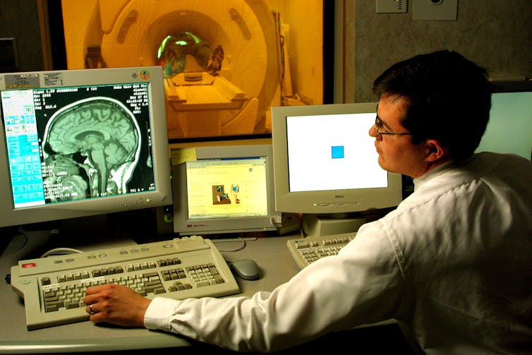
Thoughts are invisible, so what should we measure?
Thought is ultimately an internal and very individualized process that’s not readily observable. It relies on interactions across complex networks of neurons distributed throughout the peripheral and central nervous systems. Researchers can use imaging techniques, such as functional magnetic resonance imaging and electroencephalography , to see what areas of the nervous system are active during different thought processes, and how information flows through the nervous system. We’re still a long way from reliably relating these signals to the mental events they represent, though.
Many scientists consider the best proxy measure of the speed or efficiency of thought processes to be reaction time – the time from the onset of a specific signal to the moment an action is initiated. Indeed, researchers interested in assessing how fast information travels through the nervous system have used reaction time since the mid-1800s . This approach makes sense because thoughts are ultimately expressed through overt actions. Reaction time provides an index of how efficiently someone receives and interprets sensory information, decides what to do based on that information, and plans and initiates an action based on that decision.
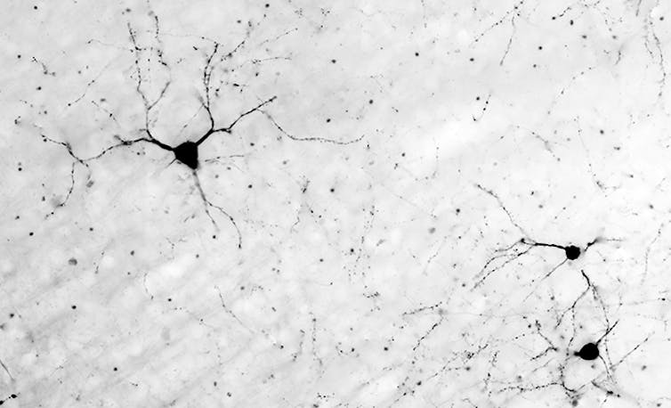
Neural factors involved
The time it takes for all thoughts to occur is ultimately shaped by the characteristics of the neurons and the networks involved. Many things influence the speed at which information flows through the system, but three key factors are:
Distance – The farther signals need to travel, the longer the reaction time is going to be. Reaction times for movements of the foot are longer than for movements of the hand, in large part because the signals traveling to and from the brain have a longer distance to cover. This principle is readily demonstrated through reflexes (note, however, that reflexes are responses that occur without “thought” because they do not involve neurons that engaged in conscious thought). The key observation for the present purpose is that the same reflexes evoked in taller individuals tend to have longer response times than for shorter individuals. By way of analogy, if two couriers driving to New York leave at the same time and travel at exactly the same speed, a courier leaving from Washington, DC will always arrive before one leaving from Los Angeles.
Neuron characteristics – The width of the neuron is important. Signals are carried more quickly in neurons with larger diameters than those that are narrower – a courier will generally travel faster on wide multi-lane highways than on narrow country roads.
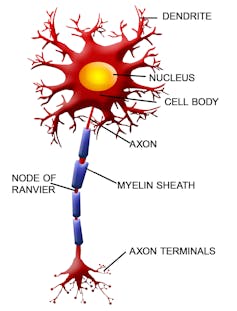
How much myelination a neuron has is also important. Some nerve cells have myelin cells that wrap around the neuron to provide a type of insulation sheath. The myelin sheath isn’t completely continuous along a neuron; there are small gaps in which the nerve cell is exposed. Nerve signals effectively jump from exposed section to exposed section instead of traveling the full extent of the neuronal surface. So signals move much faster in neurons that have myelin sheaths than in neurons that don’t. The message will get to New York sooner if it passes from cellphone tower to cellphone tower than if the courier drives the message along each and every inch of the road. In the human context, the signals carried by the large-diameter, myelinated neurons that link the spinal cord to the muscles can travel at speeds ranging from 70-120 meters per second (m/s) (156-270 miles per hour[mph]), while signals traveling along the same paths carried by the small-diameter, unmyelinated fibers of the pain receptors travel at speeds ranging from 0.5-2 m/s (1.1-4.4 mph). That’s quite a difference!
- Complexity – Increasing the number of neurons involved in a thought means a greater absolute distance the signal needs to travel – which necessarily means more time. The courier from Washington, DC will take less time to get to New York with a direct route than if she travels to Chicago and Boston along the way. Further, more neurons mean more connections. Most neurons are not in physical contact with other neurons. Instead, most signals are passed via neurotransmitter molecules that travel across the small spaces between the nerve cells called synapses. This process takes more time (at least 0.5 ms per synapse) than if the signal was continually passed within the single neuron. The message carried from Washington, DC will take less time to get to New York if one single courier does the whole route than if multiple couriers are involved, stopping and handing over the message several times along the way. In truth, even the “simplest” thoughts involve multiple structures and hundreds of thousands of neurons.

How quickly it can happen
It’s amazing to consider that a given thought can be generated and acted on in less than 150 ms. Consider the sprinter at a starting line. The reception and perception of the crack of the starter’s gun, the decision to begin running, issuing of the movement commands, and generating muscle force to start running involves a network that begins in the inner ear and travels through numerous structures of the nervous system before reaching the muscles of the legs. All that can happen in literally half the time of a blink of an eye.
Although the time to initiate a sprint start is extremely short, a variety of factors can influence it. One is the loudness of the auditory “go” signal . Although reaction time tends to decrease as the loudness of the “go” increases, there appears to be a critical point in the range of 120-124 decibels where an additional decrease of approximately 18 ms can occur. That’s because sounds this loud can generate the “startle” response and trigger a pre-planned sprinting response.
Researchers think this triggered response emerges through activation of neural centers in the brain stem . These startle-elicited responses may be quicker because they involve a relatively shorter and less complex neural system – one that does not necessarily require the signal to travel all the way up to the more complex structures of the cerebral cortex. A debate could be had here as to whether or not these triggered responses are “thoughts,” because it can be questioned whether or not a true decision to act was made; but the reaction time differences of these responses illustrate the effect of neural factors such as distance and complexity. Involuntary reflexes, too, involve shorter and simpler circuitry and tend to take less time to execute than voluntary responses.

Perceptions of our thoughts and actions
Considering how quickly they do happen, it’s little wonder we often feel our thoughts and actions are nearly instantaneous. But it turns out we’re also poor judges of when our actions actually occur.
Although we’re aware of our thoughts and the resulting movements, an interesting dissociation has been observed between the time we think we initiate a movement and when that movement actually starts . In studies, researchers ask volunteers to watch a second hand rotate around a clock face and to complete a simple rapid finger or wrist movement, such as a key press, whenever they liked. After the clock hand had completed its rotation, the people were asked to identify where the hand was on the clock face when they started their own movement.
Surprisingly, people typically judge the onset of their movement to occur 75-100 ms prior to when it actually began. This difference cannot be accounted for simply by the time it takes for the movement commands to travel from the brain to the arm muscles (which is on the order of 16-25 ms). It’s unclear exactly why this misperception occurs, but it’s generally believed that people base their judgment of movement onset on the time of the decision to act and the prediction of the upcoming movement, instead of on the movement itself. These and other findings raise important questions about the planning and control of action and our sense of agency and control in the world – because our decision to act and our perception of when we act appear to be distinct from when we in fact do.
In sum, although quantifying a single “speed of thought” may never be possible, analyzing the time it takes to plan and complete actions provides important insights into how efficiently the nervous system completes these processes, and how changes associated with movement and cognitive disorders affect the efficiency of these mental activities.
- Neuroscience
Faculty of Law - Academic Appointment Opportunities
Operations Manager
Senior Education Technologist
Audience Development Coordinator (fixed-term maternity cover)
Lecturer (Hindi-Urdu)
- The Magazine
- Stay Curious
- The Sciences
- Environment
- Planet Earth
Numbers: The Nervous System, From 268-MPH Signals to Trillions of Synapses
268 Speed (in miles per hour) at which signals travel along an alpha motor neuron in the spinal cord, the fastest such transmission in the human body. Sensory receptors in the skin, which lack the speed-boosting insulating layer called a myelin sheath, are among the slowest, at 1 mph.
100,000 Miles of myelin-covered nerve fibers in the brain of an average 20-year-old. Neuroscientists at UCLA, who have studied myelination in the brains of adults ages 23 to 80, reported in September that the coating peaks around age 39 —the same age at which participants hit top speeds in standard tests of motor abilities.
100 trillion Minimum number of neural connections, or synapses, in the human brain. That is at least 1,000 times the number of stars in our galaxy. British researchers reported in December that genes involved in the workings of synapses account for about 7 percent of our genome.
50 Depth, in nanometers, of the smallest grooves detectable by a human fingertip (that is about 2 millionths of an inch). Most of the 2 billion or so nerve endings in the outermost layer of our skin sense pain; those dedicated to temperature allow us to detect differences as small as 0.01 degree Fahrenheit.
2,000 Number of slices created from the cerebral cortex of a mouse by Harvard University scientists . The researchers will image each slice under an electron microscope and then build a 3-D picture of all of the brain’s connections. Someday, similar maps of human brains may yield clues to mental illness, memory, and personality traits.
1 billion Number of neurons, linked by 10 trillion synapses, in a brain simulation developed by IBM and Lawrence Berkeley National Lab , running on the Dawn supercomputer. Researchers are testing hypotheses about how the brain works. The real human brain contains about 100 billion neurons, so scientists are getting close—in raw numbers, at least.
- neuroscience
- brain structure & function
Already a subscriber?
Register or Log In

Keep reading for as low as $1.99!
Sign up for our weekly science updates.
Save up to 40% off the cover price when you subscribe to Discover magazine.


How does a nerve impulse travel through the body?
The information acquired at the dendritic tip of a nerve cell sets off a chemical reaction that creates an electrical impulse. this impulse travels from the dendrite to the cell body and then along the axon to its end. at the end of the axon the electrical impulse sets off the release of some chemicals. these chemicals cross the gap or synapse and start a similar electrical impulse in a dendrite of the next neuron. a similar synapse finally allows the delivery of such impulse from neurons to other cells such as muscles or glands..

define nerve impulse which structure in a neuron help to conduct a nerve impulse :
i} towards the cell
ii} away from the cell body
What are the scheme of travelling of nerve impulse in the body?

IMAGES
VIDEO
COMMENTS
A nerve impulse causes Na + to enter the cell, resulting in (b) depolarization. At the peak action potential, ... For an action potential to communicate information to another neuron, it must travel along the axon and reach the axon terminals where it can initiate neurotransmitter release. The speed of conduction of an action potential along an ...
The transmission of a nerve impulse along a neuron from one end to the other occurs as a result of electrical changes across the membrane of the neuron. The membrane of an unstimulated neuron is polarized—that is, there is a difference in electrical charge between the outside and inside of the membrane. The inside is negative with respect to ...
A nerve impulse is an all-or-nothing response depending on if the stimulus input was strong enough to reach threshold. If a neuron responds at all, it responds completely. A greater stimulation does not produce a stronger impulse. Figure 8.4.2 An action potential speeds along an axon in milliseconds.
Like the heart, lungs, and stomach, the nervous system is made up of specialized cells. These include nerve cells (or neurons) and glial cells (or glia ). Neurons are the basic functional units of the nervous system, and they generate electrical signals called action potentials, which allow them to quickly transmit information over long distances.
8.4 Summary. A nerve impulse is an electrical phenomenon that occurs because of a difference in electrical charge across the plasma membrane of a neuron. The sodium-potassium pump maintains an electrical gradient across the plasma membrane of a neuron when it is not actively transmitting a nerve impulse. This gradient is called the resting ...
Neurotransmitters are how we communicate between one cell and the next. Synapses between neurons are either excitatory or inhibitory - and that all comes down to the neurotransmitter released. Excitatory neurotransmitters cause the signal to propagate - more action potentials are triggered. Inhibitory signals work to cancel the signal.
At a synapse, one neuron sends a message to a target neuron—another cell. Most synapses are chemical; these synapses communicate using chemical messengers. Other synapses are electrical; in these synapses, ions flow directly between cells. At a chemical synapse, an action potential triggers the presynaptic neuron to release neurotransmitters.
Transmission of Nerve Impulses. Neurones transmit electrical impulses, which travel extremely quickly along the neurone cell surface membrane from one end of the neurone to the other. Unlike a normal electric current, these impulses are not a flow of electrons. These impulses, known as action potentials, occur via very brief changes in the ...
A nerve impulse is the electric signals that pass along the dendrites to generate a nerve impulse or an action potential. An action potential is due to the movement of ions in and out of the cell. It specifically involves sodium and potassium ions. They are moved in and out of the cell through sodium and potassium channels and sodium-potassium ...
Well, in the amount of time it takes you to say "Mississippi" three times, a nerve could send an electrical signal the length of a football field and back. That is fast! Now that you know how fast a nerve impulse can travel, let's take a look at the system that is the highway for these electrical signals.
Nerve impulse propagates by jumping from one node of Ranvier to the next. This makes the process of nerve impulse faster as the nerve impulse does not travel the entire length of the axon ( this happens in the case of continuous conduction). The nerve impulse travels at a speed of 100 m/s in saltatory conduction.
Nerve impulses travel much faster in nerves with a myelin sheath than in those without one. Insulating a Nerve Fiber. Most nerve fibers inside and outside the brain are wrapped with many layers of tissue composed of a fat (lipoprotein) called myelin. These layers form the myelin sheath. Much like the insulation around an electrical wire, the ...
In the human context, the signals carried by the large-diameter, myelinated neurons that link the spinal cord to the muscles can travel at speeds ranging from 70-120 meters per second (m/s) (156 ...
268 Speed (in miles per hour) at which signals travel along an alpha motor neuron in the spinal cord, the fastest such transmission in the human body. Sensory receptors in the skin, which lack the speed-boosting insulating layer called a myelin sheath, are among the slowest, at 1 mph. 100,000 Miles of myelin-covered nerve fibers in the brain of an average 20-year-old.
Nerve conduction velocity. Saltatory conduction. In neuroscience, nerve conduction velocity ( CV) is the speed at which an electrochemical impulse propagates down a neural pathway. Conduction velocities are affected by a wide array of factors, which include age, sex, and various medical conditions. Studies allow for better diagnoses of various ...
Nerve impulses, also known as action potentials, travel at different speeds depending on the type of nerve fiber. In general, they can travel anywhere from 0.5 to 120 meters per second (m/s). Myelinated nerve fibers, which have a protective sheath, conduct impulses faster than unmyelinated fibers. The speed of nerve impulses also depends on the diameter of the nerve fiber, with larger diameter ...
The information acquired at the dendritic tip of a nerve cell sets off a chemical reaction that creates an electrical impulse. This impulse travels from the dendrite to the cell body and then along the axon to its end. At the end of the axon the electrical impulse sets off the release of some chemicals.